Alternate header for print version
Advanced search
Contributors
Help
Submit
Search
menu
Cell Process
Cell Component
Cell Type
Organism
Microbial
Alzheimer's
Data Sets
Center for Research in Biological Systems
University of California, San Diego
9500 Gilman Drive
La Jolla, CA 92093-0608, USA
Voice
: (858) 534-0276
Fax
: (858) 534-7497
Email
: dorloff@ncmir.ucsd.edu
Search Results for
laser scanning confocal
(395 results)
(Not the results you were expecting?)
(Comments)
Still Images
Video/Animation
Z-Stack
Time Series
CIL:12292
NCBI Organism Classification
Mus musculus
Biological Process
cell-substrate adhesion
Cellular Component
microtubule
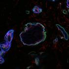
Colocalization of the microtubule anchoring factor LL5α (red) with integrin α6 (blue) in the basal cortex of outer epithelium of mouse mammary gland tissue, and laminin (green) in the basement membr...
CIL:35290
NCBI Organism Classification
Lilium formosanum
Biological Process
pollen tube growth
Cellular Component
microtubule
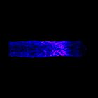
Freeze substituted Lilium pollen tube immunolabeled for microtubules (blue>magenta) and imaged using laser scanning confocal microscopy. Shown is a projection of x-y slices revealing the distribution ...
CIL:12294
NCBI Organism Classification
Homo sapiens
Biological Process
cell-substrate adhesion
Cellular Component
microtubule
Localization of the microtubule anchoring factor LL5α (red), microtubules (green), and the microtubule plus-end binding protein, EB1 (blue) in an MCF-10Aeco epithelial cell. EB1 was concentrated in ...
CIL:35293
NCBI Organism Classification
Lilium formosanum
Biological Process
pollen tube growth
Cellular Component
microtubule
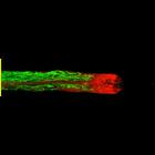
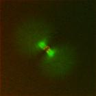
Living Lilium pollen tube labeled with paclitaxel (blue) to mark microtubules (blue) and cytochalasin D TMR to mark endoplasmic reticulum (green) imaged using laser scanning focal microscopy. The grow...
CIL:35294
NCBI Organism Classification
Lilium formosanum
Biological Process
pollen tube growth
Cellular Component
mitochondria
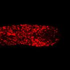
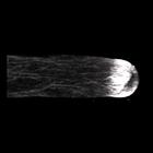
Living Lilium pollen tube labeled with TMRE (red)to mark mitochondria and imaged using laser scanning focal microscopy. Shown is a central 1.0 micron thick x-y slice. The growing tip is devoid of mit...
CIL:35295
NCBI Organism Classification
Lilium formosanum
Biological Process
pollen tube growth
Cellular Component
plant-type vacuole lumen
Living Lilium pollen tube labeled with carboxy-dichlorofluorofluorescein (green) to mark the vacuole and cytochalasin D TMR to mark endoplasmic reticulum (red) imaged using laser scanning focal micros...
CIL:35158
NCBI Organism Classification
Homo sapiens
Biological Process
mitosis
Cellular Component
mitotic membranes
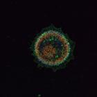
Live mitotic HeLa cell treated with epsin1 siRNA, DiOC6(3)to label mitotic membranes (green), and Hoechst 33258 to label chromosomes (blue). Confocal images were taken at 0.118 μm steps along the Z-...
CIL:12290
NCBI Organism Classification
Homo sapiens
Biological Process
cell-substrate adhesion
Cellular Component
microtubule
To determine the extent to which extracellular matrix proteins colocalize with the microtubule-anchoring factor LL5s (LL5α), MCF-10A epithelial cells were immunostained with antibodies to laminin rec...
CIL:36572
NCBI Organism Classification
Xenopus laevis
Biological Process
first cell cycle
Cellular Component
microtubule
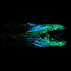
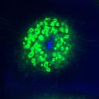
This image is part of a large data set of Xenopus laevis eggs imaged at various times post fertilization (the first number of the file name corresponds minutes, eg 30_2 is 30 min post fertilization). ...
CIL:35285
NCBI Organism Classification
Lilium formosanum
Biological Process
pollen tube growth
Cellular Component
actin cytoskeleton
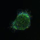
Freeze substituted Lilium pollen tube immunolabeled for filamentous actin and imaged using laser scanning confocal microscopy. Shown is a projection of z-slices revealing the dense sub-apical actin fr...
« Previous
1
2
3
4
5
6
7
8
9
...
40
Next »
Results per page:
10
20
50
100