Licensing

Pd
Public Domain: This image is in the public domain and thus free of any copyright restrictions. However, as is the norm in scientific publishing and as a matter of courtesy, any user should credit the content provider for any public or private use of this image whenever possible. Learn more
Comments: Powered by Pnyxe

Description

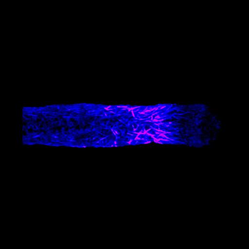
Freeze substituted Lilium pollen tube immunolabeled for microtubules (blue>magenta) and imaged using laser scanning confocal microscopy. Shown is a projection of x-y slices revealing the distribution of microtubules in the sub-apical region. This group of images also contains an x-z projection of the same specimen.

Technical Details

Germinated lily pollen on agar loops was plunge frozen, freeze substituted, labeled with an anti-tubulin mouse monoclonal antibody followed by Cy-3 goat anti-mouse and imaged with a Zeiss 510 meta confocal microscope using a 63x 1.4 NA objective lens. The image shown is a projection of x-y slices. See Lovy-Wheeler et al. 2005, Planta 221:95-104.

Biological Sources

NCBI Organism Classification
Lilium formosanum
(St Joseph's lily)
Cell Type
pollen tube
Cellular Component
microtubule

Biological Context

Biological Process
pollen tube growth

Attribution

Names
Alenka Lovy-Wheeler
Kathleen L Wilsen
Tobias I Baskin
Peter K Hepler

Grouping

This image is part of a group.

Imaging

Image Type
photomultiplier tube (PMT)
Imaging Mode
confocal microscopy
Parameters Imaged
distribution of epitope
Source of Contrast
distribution of a specific protein
Visualization Methods
Cy3
Processing History
projection of confocal slices
Data Qualifiers
processed data
suitable for spatial measurements

Sample Preparation

Methods
cryofixed tissue
FBbi:00000621
Relation To Intact Cell
whole mounted tissue

Dimensions

Spatial Axis Image Size Pixel Size
X 2001px ——
Y 424px ——
*CIL – Cell Image Library accession number. Please use this to reference an image.