Alternate header for print version
Advanced search
Contributors
Help
Submit
Search
menu
Cell Process
Cell Component
Cell Type
Organism
Microbial
Alzheimer's
Data Sets
Center for Research in Biological Systems
University of California, San Diego
9500 Gilman Drive
La Jolla, CA 92093-0608, USA
Voice
: (858) 534-0276
Fax
: (858) 534-7497
Email
: dorloff@ncmir.ucsd.edu
Search Results for
laser scanning confocal
(395 results)
(Not the results you were expecting?)
(Comments)
Still Images
Video/Animation
Z-Stack
Time Series
CIL:35286
NCBI Organism Classification
Lilium formosanum
Biological Process
pollen tube growth
Cellular Component
actin cytoskeleton
Freeze substituted Lilium pollen tube immunolabeled for filamentous actin and imaged using laser scanning confocal microscopy. Shown is a projection of x-z slices revealing the cortical distribution o...
CIL:35291
NCBI Organism Classification
Lilium formosanum
Biological Process
pollen tube growth
Cellular Component
microtubule
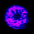
Freeze substituted Lilium pollen tube immunolabeled for microtubules (blue>magenta) and imaged using laser scanning confocal microscopy. Shown is a projection of x-z slices revealing the cortical dist...
CIL:35161
NCBI Organism Classification
Homo sapiens
Biological Process
none specified
Cellular Component
mitotic membranes
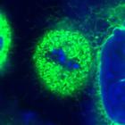
Live mitotic HeLa cell treated with control siRNA, DiOC6(3)to label mitotic membranes (green), and Hoechst 33258 to label chromosomes (blue). Confocal images were taken at 0.118 μm steps along the Z-...
CIL:48305
NCBI Organism Classification
Sprague-Dawley rat
Biological Process
none specified
Cellular Component
atrial cells
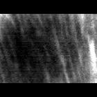
Images are acquired from fixed and living heart tissue using fiber-optics and laser-scanning confocal microscopy, respectively. Three sets of images denoted as CCM, FCMtopical, FCMcarrier were stored ...
CIL:48306
NCBI Organism Classification
Sprague-Dawley rat
Biological Process
none specified
Cellular Component
nodal cells
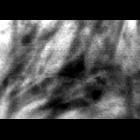
Images are acquired from fixed and living heart tissue using fiber-optics and laser-scanning confocal microscopy, respectively. Three sets of images denoted as CCM, FCMtopical, FCMcarrier were stored ...
CIL:35563
NCBI Organism Classification
Rattus rattus
Biological Process
cell proliferation
Cellular Component
keratin filament
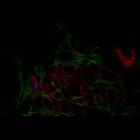
Liver of a rat exposed to chemical carcinogens. Serial sections of the liver imaged by laser scanning confocal microscopy. F-actin (red) is in bile canaliculi at junctions of the hepatocytes, which ...
CCDB:44959
Species
Rat
Organ
brain
Cell type
astrocyte
System
Central nervous system
Structure
None
Diatheva Panx1 Ab in Rat Cerebellum
CIL:35289
NCBI Organism Classification
Lilium formosanum
Biological Process
pollen tube growth
Cellular Component
endoplasmic reticulum
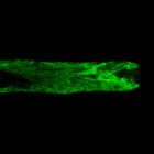
Living Lilium pollen tube labeled with mGFP5-HDEL to mark endoplasmic reticulum (green) imaged using laser scanning focal microscopy. Shown is a central 1.0 micron thick x-y slice. The growing tip is...
CIL:12288
NCBI Organism Classification
Homo sapiens
Biological Process
cell-substrate adhesion
Cellular Component
microtubule
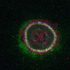
To determine the extent to which extracellular matrix proteins colocalize with the microtubule-anchoring factor LL5s (LL5α), MCF-10A epithelial cells were immunostained with antibodies to the secrete...
CIL:13402
NCBI Organism Classification
Strongylocentrotus purpuratus
Biological Process
mitotic anaphase
Cellular Component
microtubule
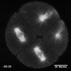
Eight-cell purple urchin embryo expressing three tandem GFPs fused to the ensconsin microtubule-binding domain (EMTB-3G; Faire et al., 1999). The embryo was treated with 20 µM nocodazole at time 00:...
« Previous
1
...
3
4
5
6
7
8
9
10
...
40
Next »
Results per page:
10
20
50
100