Licensing

Pd
Public Domain: This image is in the public domain and thus free of any copyright restrictions. However, as is the norm in scientific publishing and as a matter of courtesy, any user should credit the content provider for any public or private use of this image whenever possible. Learn more
Comments: Powered by Pnyxe

Description

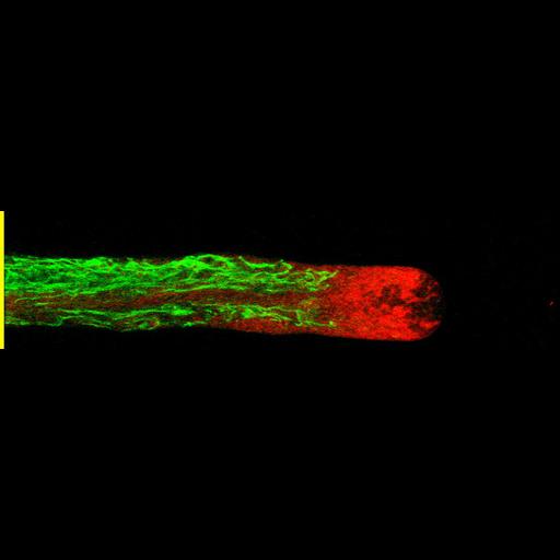
Living Lilium pollen tube labeled with carboxy-dichlorofluorofluorescein (green) to mark the vacuole and cytochalasin D TMR to mark endoplasmic reticulum (red) imaged using laser scanning focal microscopy. Shown is a central 1.0 micron thick x-y slice. The growing tip is devoid of both organelles. Other images in the group reveal the distributions of additional cytoplasmic components.

Technical Details

Living lily pollen tubes were labeled and then imaged with a Zeiss 510 meta confocal microscope using a 63x 1.4 NA objective lens. Green and red signals from a central plane through the tube were merged to generate the image. See Lovy-Wheeler et al. 2007 Cell Motil Cytoskel 64:217-321.

Biological Sources

NCBI Organism Classification
Lilium formosanum
(St Joseph's lily)
Cell Type
pollen tube
Cellular Component
plant-type vacuole lumen
endoplasmic reticulum

Biological Context

Biological Process
pollen tube growth

Attribution

Names
Alenka Lovy-Wheeler
Luis Cardenas
Joseph Kunkel
Peter K Hepler

Grouping

This image is part of a group.

Imaging

Image Type
photomultiplier tube (PMT)
Imaging Mode
confocal microscopy
Parameters Imaged
fluorescence emission
Source of Contrast
compartmentalization of stain or label
Visualization Methods
carboxy-DCFDA
Tetramethyl rhodamine (TRITC)
Processing History
merged confocal slices
Data Qualifiers
processed data
suitable for spatial measurements

Sample Preparation

Methods
living tissue
Relation To Intact Cell
whole mounted tissue

Dimensions

Spatial Axis Image Size Pixel Size
X 983px ——
Y 242px ——
*CIL – Cell Image Library accession number. Please use this to reference an image.