Licensing

Pd
Public Domain: This image is in the public domain and thus free of any copyright restrictions. However, as is the norm in scientific publishing and as a matter of courtesy, any user should credit the content provider for any public or private use of this image whenever possible. Learn more
Comments: Powered by Pnyxe

Description

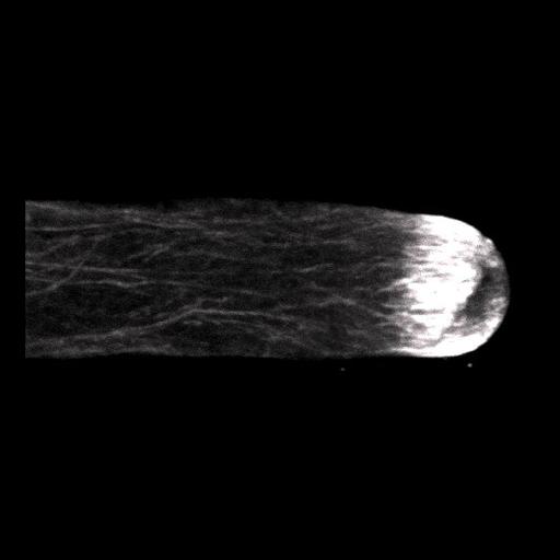
Freeze substituted Lilium pollen tube immunolabeled for filamentous actin and imaged using laser scanning confocal microscopy. Shown is a projection of z-slices revealing the dense sub-apical actin fringe. Other images in the group reveal the distributions of other cytoskeletal elements and membranous components.

Technical Details

Germinated lily pollen on agar loops was plunge frozen, freeze substituted, labeled with an anti-actin mouse monoclonal antibody followed by Cy-3 goat anti-mouse and imaged with a Zeiss 510 meta confocal microscope using a 63x 1.4 NA objective lens. The image shown is a projection of z-slices. See Lovy-Wheeler et al. 2005, Planta 221:95-104.

Biological Sources

NCBI Organism Classification
Lilium formosanum
(St Joseph's lily)
Cell Type
pollen tube
Cellular Component
actin cytoskeleton

Biological Context

Biological Process
pollen tube growth

Attribution

Names
Alenka Lovy-Wheeler
Kathleen L Wilsen
Tobias I Baskin
Peter K Hepler

Grouping

This image is part of a group.

Imaging

Image Type
photomultiplier tube (PMT)
Imaging Mode
confocal microscopy
Parameters Imaged
fluorescence emission
Source of Contrast
differences in adsorption or binding of stain
Visualization Methods
Cy3
Processing History
projection of confocal slices
Data Qualifiers
processed data
suitable for spatial measurements

Sample Preparation

Methods
cryofixed tissue
FBbi:00000621
Relation To Intact Cell
whole mounted tissue

Dimensions

Spatial Axis Image Size Pixel Size
X 532px ——
Y 216px ——
*CIL – Cell Image Library accession number. Please use this to reference an image.