Alternate header for print version
Advanced search
Contributors
Help
Submit
Search
menu
Cell Process
Cell Component
Cell Type
Organism
Microbial
Alzheimer's
Data Sets
Center for Research in Biological Systems
University of California, San Diego
9500 Gilman Drive
La Jolla, CA 92093-0608, USA
Voice
: (858) 534-0276
Fax
: (858) 534-7497
Email
: dorloff@ncmir.ucsd.edu
Search Results for
confocal
(3462 results)
(Not the results you were expecting?)
(Comments)
Still Images
Video/Animation
Z-Stack
Time Series
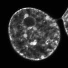
CIL:1315
NCBI Organism Classification
Mus musculus
Biological Process
nucleus organization
Cellular Component
nucleus
The intracellular mobility of MeCP2 (a methylated DNA-binding protein) followed in a mouse fibroblast stably expressing GFP-MeCP2 through a time series of images captured by confocal microscopy after ...
CIL:132
NCBI Organism Classification
Mus musculus
Biological Process
regulation of actin cytoskeleton organization
Cellular Component
cell leading edge
NIH 3T3 cell transfected with EGFP-VASP. VASP is localized to the focal adhesions and is also present along the protruding leading edge. VASP only hightlights the portions of the periphery that are ...
CIL:35293
NCBI Organism Classification
Lilium formosanum
Biological Process
pollen tube growth
Cellular Component
microtubule
Living Lilium pollen tube labeled with paclitaxel (blue) to mark microtubules (blue) and cytochalasin D TMR to mark endoplasmic reticulum (green) imaged using laser scanning focal microscopy. The grow...
CIL:35294
NCBI Organism Classification
Lilium formosanum
Biological Process
pollen tube growth
Cellular Component
mitochondria
Living Lilium pollen tube labeled with TMRE (red)to mark mitochondria and imaged using laser scanning focal microscopy. Shown is a central 1.0 micron thick x-y slice. The growing tip is devoid of mit...
CIL:35295
NCBI Organism Classification
Lilium formosanum
Biological Process
pollen tube growth
Cellular Component
plant-type vacuole lumen
Living Lilium pollen tube labeled with carboxy-dichlorofluorofluorescein (green) to mark the vacuole and cytochalasin D TMR to mark endoplasmic reticulum (red) imaged using laser scanning focal micros...
CIL:35158
NCBI Organism Classification
Homo sapiens
Biological Process
mitosis
Cellular Component
mitotic membranes
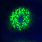
Live mitotic HeLa cell treated with epsin1 siRNA, DiOC6(3)to label mitotic membranes (green), and Hoechst 33258 to label chromosomes (blue). Confocal images were taken at 0.118 μm steps along the Z-...
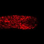
CIL:40252
NCBI Organism Classification
Rattus
Biological Process
forebrain astrocyte development
Cellular Component
cell projection cytoplasm
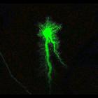
Intracellular dye injections of protoplasmic astrocytes from the CA1 region of hippocampus of a 1 month old rat reveals highly ramified spongiform processes that span territories with minimal overlap....
CIL:40658
NCBI Organism Classification
Drosophila melanogaster
Biological Process
microtubule cytoskeleton organization
Cellular Component
microtubule
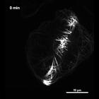
Time series spinning disc confocal images of Drosophila S2 cells stably expressing pMT-Eos2-alpha tubulin. A diagonal ~2.5 micrometer strip was photoconverted and images taken every minute for 20 minu...
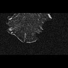
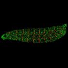
CIL:41566
NCBI Organism Classification
Claviceps
Biological Process
fungal infection
Cellular Component
hyphae
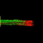
This confocal micrograph shows wheat stigma (green) infected with Claviceps fungus (yellow). The image was captured with a confocal microscope using a 40X objective. A related image is available as ...
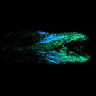
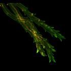
CIL:41824
NCBI Organism Classification
Drosophila melanogaster
Biological Process
sensory neuron organization
Cellular Component
neuron projection
Confocal image stacks of Drosophila (fruit fly) larva with labeled dendrite arborization sensory neurons and epidermal cells. Green is dendrites of sensory neurons on the body wall and red is attachme...
« Previous
1
...
4
5
6
7
8
9
10
11
...
347
Next »
Results per page:
10
20
50
100