Alternate header for print version


Licensing
Public Domain: This image is in the public domain and thus free of any copyright restrictions. However, as is the norm in scientific publishing and as a matter of courtesy, any user should credit the content provider for any public or private use of this image whenever possible. Learn more
tweet  
*CIL – Cell Image Library accession number. Please use this to reference an image.

CIL:132*  Cite 
Description

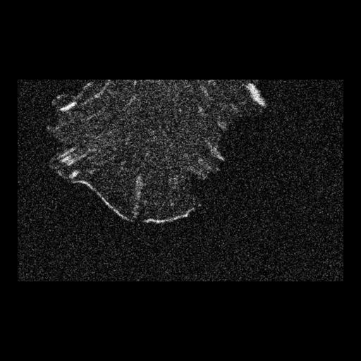
NIH 3T3 cell transfected with EGFP-VASP. VASP is localized to the focal adhesions and is also present along the protruding leading edge. VASP only hightlights the portions of the periphery that are advancing forward, giving VASP a non-uniform distribution along the leading edge. Time series taken as a single confocal slice using a Zeiss LSM 510 confocal with a 100X 1.3 NA objective, 488 nm excitation and a 505-530 BP emission filter.

Biological Sources
NCBI Organism Classification
Mus musculus
Cell Type
fibroblast
Cell Line
NIH/3T3
Cellular Component
cell leading edge
focal adhesion
cytoskeleton
Biological Context
Biological Process
regulation of actin cytoskeleton organization
Molecular Function
structural constituent of cytoskeleton
Attribution
Names
Catherine Galbraith,
Citation
Digital Object Identifier (DOI)
doi:10.7295/W9CIL132
Archival Resource Key (ARK)
ark:/b7295/w9cil132
Imaging
Image Type
recorded image
Image Mode
single-spot confocal microscopy
Parameters Imaged
fluorescence emission
Source of Contrast
distribution of a specific protein
Visualization Methods
EGFP
Processing History
non-linear methods
Sample Preparation
Methods
living tissue
Relation To Intact Cell
living tissue
Dimensions
Spatial Axis Image Size Pixel Size
X 462px 0.04µm
Y 286px 0.04µm