Alternate header for print version
Advanced search
Contributors
Help
Submit
Search
menu
Cell Process
Cell Component
Cell Type
Organism
Microbial
Alzheimer's
Data Sets
Center for Research in Biological Systems
University of California, San Diego
9500 Gilman Drive
La Jolla, CA 92093-0608, USA
Voice
: (858) 534-0276
Fax
: (858) 534-7497
Email
: dorloff@ncmir.ucsd.edu
Search Results for
James Jamieson
(124 results)
(Not the results you were expecting?)
(Comments)
Still Images
Video/Animation
Z-Stack
Time Series
CIL:37195
NCBI Organism Classification
Neurospora
Biological Process
none specified
Cellular Component
mitochondrion
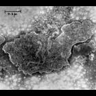
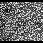
Transmission electron micrograph of a negatively stained mitochondria from the fungi Neurospora. The use of osmium tetroxide as a fixative for electron microscopy was first described by Dr. Palade at ...
CIL:37218
NCBI Organism Classification
Rattus
Biological Process
transmission of nerve impulse
Cellular Component
myelin sheath
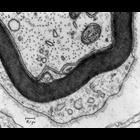
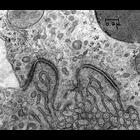
Transmission electron micrograph of a myelinated axon from a rat tongue. Image made available by James D. Jamieson and the Department of Cell Biology, Yale University School of Medicine.
CIL:37221
NCBI Organism Classification
none specified
Biological Process
none specified
Cellular Component
Nissl body
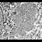
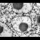
Transmission electron micrograph of the accumulation of rough endoplasmic reticulum known as Nissl bodies within nerve cells from the superior cervical ganglion. Note that this very early (1953) mic...
CIL:37223
NCBI Organism Classification
none specified
Biological Process
cytoplasm organization
Cellular Component
Nissl body
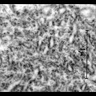
Transmission electron micrograph showing the aggregation of rough endplasmic reticulum known as a Nissl bodiy within nerve cells of the superior cervical ganglion. Note that this very early (1953) m...
CIL:37234
NCBI Organism Classification
Cavia porcellus
Biological Process
enzyme seretion
Cellular Component
rough endoplasmic reticulum
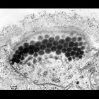
Early transmission electron micrograph of guinea pig pancreas showing ribosome-studded rough endoplasmic reticulum with secretory granules accumulating in the lumen (cisternae). Image made available ...
CIL:37250
NCBI Organism Classification
Chlamydomonas
Biological Process
organelle organization
Cellular Component
chloroplast
An electron micrograph of a potion of a cell of the unicellular alga Chlamydomonas reinhardii illustrating the eye spot, an organelle found inside the chloroplast that is composed of red carotenoid pi...
CIL:37260
NCBI Organism Classification
Chlamydomonas reinhardtii
Biological Process
flagellum organization
Cellular Component
flagellum
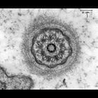
An electron micrograph of a cross section of a flagellum from the unicellular alga Chlamydomonas (Yellow strain) showing the characteristic 9-fold symmetry. This section, close to the basal body does ...
CIL:37263
NCBI Organism Classification
Bos taurus
Biological Process
none specified
Cellular Component
zymogen granule
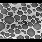
Zymogen granule fraction Calf. Zymogen granules show their homogeneous content and limiting membrane. Image made available by James D. Jamieson and the Department of Cell Biology, Yale University S...
CIL:37264
NCBI Organism Classification
Bos taurus
Biological Process
none specified
Cellular Component
zymogen granule
Zymogen granule fraction Calf. Section through a pellet of a zymogen granule fraction showing that the preparation mostly consists of morphologically unaltered zymogen granules. Image made available...
CIL:37168
NCBI Organism Classification
Rattus
Biological Process
adherens junction organization
Cellular Component
adherens junction
Transmission electron micrograph of adherens junctions (macula adherens or desmosome)in the proximal tubule of a rat kidney. The junctions with associated tonofilaments are seen at the apposed plasma...
« Previous
1
2
3
4
5
6
7
8
9
...
13
Next »
Results per page:
10
20
50
100