Licensing

Attribution_nc_sa
Attribution Non-Commercial Share Alike:This image is licensed under a Creative Commons Attribution, Non-Commercial Share Alike License. View License Deed | View Legal Code
Comments: Powered by Pnyxe

Description

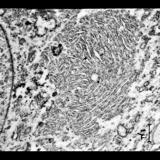
Transmission electron micrograph of the accumulation of rough endoplasmic reticulum known as Nissl bodies within nerve cells from the superior cervical ganglion. Note that this very early (1953) micrograph is primarily of historical interest since it pre-dates glutaraldehye fixation which was introduced in the early 60's and is considered essential for adequate nerve tissue preservation. Examples of nerve tissue micrographs prepared in 1965 are CIL:37218, CIL:37219 and CIL:37220. Image made available by James D. Jamieson and the Department of Cell Biology, Yale University School of Medicine.

Technical Details

Additional reference: Palay, S and Palade, GE J Biophys Biochem Cytol. 1955 Jan;1(1):69-88. Original 3.25 in. x 4 in. lantern slides were scanned at 600dpi. Original magnification x7,600.

Biological Sources

Cell Type
neuron
Cellular Component
Nissl body
rough endoplasmic reticulum

Biological Context

Human Development Anatomy
superior cervical ganglion

Attribution

Link
George E. Palade EM Slide Collection
Date
05/07/1953
Other
Sanford Palay

Grouping

This image is part of a group.

Imaging

Image Type
recorded image
Imaging Mode
transmission electron microscopy (TEM)
Parameters Imaged
electron density
Source of Contrast
differences in adsorption or binding of stain
Visualization Methods
osmium tetroxide
Processing History
unprocessed raw data

Sample Preparation

Methods
chemically fixed tissue
Relation To Intact Cell
sectioned tissue

Dimensions

Spatial Axis Image Size Pixel Size
X 6000px ——
Y 5358px ——
*CIL – Cell Image Library accession number. Please use this to reference an image.