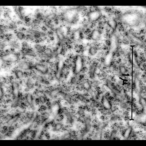
Transmission electron micrograph showing the aggregation of rough endplasmic reticulum known as a Nissl bodiy within nerve cells of the superior cervical ganglion. Note that this very early (1953) micrograph is primarily of historical interest since it pre-dates glutaraldehye fixation which was introduced in the early 60's and is considered essential for adequate nerve tissue preservation. Examples of nerve tissue micrographs prepared in 1965 are CIL:37218, CIL:37219 and CIL:37220. Image made available by James D. Jamieson and the Department of Cell Biology, Yale University School of Medicine.
Additional reference: Palay, S and Palade, GE J Biophys Biochem Cytol. 1955 Jan;1(1):69-88. Original 3.25 in. x 4 in. lantern slides were scanned at 600dpi. Original magnification 37,500x.
| Spatial Axis | Image Size | Pixel Size |
|---|---|---|
| X | 6000px | —— |
| Y | 5526px | —— |