Alternate header for print version
Advanced search
Contributors
Help
Submit
Search
menu
Cell Process
Cell Component
Cell Type
Organism
Microbial
Alzheimer's
Data Sets
Center for Research in Biological Systems
University of California, San Diego
9500 Gilman Drive
La Jolla, CA 92093-0608, USA
Voice
: (858) 534-0276
Fax
: (858) 534-7497
Email
: dorloff@ncmir.ucsd.edu
Search Results for
cortex
(640 results)
(Not the results you were expecting?)
(Comments)
Still Images
Video/Animation
Z-Stack
Time Series
CIL:54684
NCBI Organism Classification
Drosophila melanogaster
Biological Process
Glial-Glial Tiling
Cellular Component
Astrocyte membranes
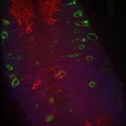
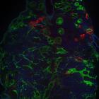
Confocal image of the CNS of a D. melanogaster in the third instar larval stage after αSNAP KD, 32uM from the ventral surface, depicting astrocytes (red), cortex glia (green) and neuronal nuclei (bl...
CIL:54660
NCBI Organism Classification
Drosophila melanogaster
Biological Process
Glial-Glial Tiling
Cellular Component
Astrocyte membranes
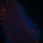
Confocal image of the CNS of a control D. melanogaster in the third instar larval stage, 70uM from the ventral surface, depicting astrocytes (red), cortex glia (green) and neuronal nuclei (blue). Our ...
CIL:54653
NCBI Organism Classification
Drosophila melanogaster
Biological Process
Glial-Glial Tiling
Cellular Component
Astrocyte membranes
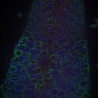
Confocal image of the CNS of a control D. melanogaster in the third instar larval stage, 0uM from the ventral surface, depicting astrocytes (red), cortex glia (green) and neuronal nuclei (blue). Our s...
CIL:54651
NCBI Organism Classification
Drosophila melanogaster
Biological Process
Glial-Glial Tiling
Cellular Component
Astrocyte membranes
Confocal image of the CNS of a control D. melanogaster in the third instar larval stage, 0uM from the ventral surface, depicting astrocytes (red), cortex glia (green) and neuronal nuclei (blue). Our s...
CIL:54663
NCBI Organism Classification
Drosophila melanogaster
Biological Process
Glial-Glial Tiling
Cellular Component
Astrocyte membranes
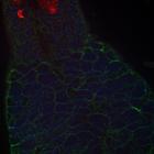
Confocal image of the CNS of a D. melanogaster in the third instar larval stage after Spz3 KD, 0uM from the ventral surface, depicting astrocytes (red), cortex glia (green) and neuronal nuclei (blue)....
CIL:39070
NCBI Organism Classification
Mus musculus
Biological Process
renal system process
Cellular Component
mitochondrion
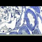
This light micrograph shows a transverse section of a region of mouse kidney cortex stained with toluidine blue. The kidney is made up of two distinct tissue regions: the medulla and the cortex. The n...
CIL:39340
NCBI Organism Classification
Nicotiana alata
Biological Process
plant stem organization
Cellular Component
none specified
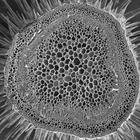
Scanning electron microscope image of Nicotiana alata stem cross section. Image shows outer epidermal layer, followed by the cortex and then large vascular bundles. The vascular bundles contain the ph...
CIL:40189
NCBI Organism Classification
Homo sapiens
Biological Process
none specified
Cellular Component
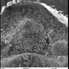
neuronal cell body
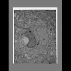
Single image from a serial section electron microscopic reconstruction of the cell body of a cortical pyramidal neuron from a biopsy sample obtained from the cortex of a patient with Alzheimer's dise...
CIL:41403
NCBI Organism Classification
Mus musculus
Biological Process
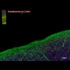
somatosensory cortex morphogenesis
Cellular Component
microtubule cytoskeleton
Video of an array tomogram of a mouse somatosensory cortex. The volume is a "fly-through" of a sagitally oriented rectangular slab of transgenic mouse somatosensory cortex. The overlay thumbnail at up...
CIL:40389
NCBI Organism Classification
Helianthus annuus
Biological Process
plant stem organization
Cellular Component
cortex
Scanning electron microscope image of cross-section through Helianthus annuus (sunflower) stem. The image shows the outer epidermal layer, followed by the cortex and then large vascular bundles. The v...
« Previous
1
2
3
4
5
6
7
8
9
...
64
Next »
Results per page:
10
20
50
100