Alternate header for print version
Advanced search
Contributors
Help
Submit
Search
menu
Cell Process
Cell Component
Cell Type
Organism
Microbial
Alzheimer's
Data Sets
Center for Research in Biological Systems
University of California, San Diego
9500 Gilman Drive
La Jolla, CA 92093-0608, USA
Voice
: (858) 534-0276
Fax
: (858) 534-7497
Email
: dorloff@ncmir.ucsd.edu
Search Results for
Drosophila S2
(27 results)
(Not the results you were expecting?)
(Comments)
Still Images
Video/Animation
Z-Stack
Time Series
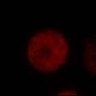
CIL:40668
NCBI Organism Classification
Drosophila melanogaster
Biological Process
microtubule cytoskeleton organization
Cellular Component
microtubule
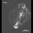
Time lapse movie prepared from spinning disc confocal images of Drosophila S2 cells stably expressing pMT-Eos2-alpha tubulin. A diagonal ~2.5 micrometer strip was photoconverted and images taken every...
CIL:36147
NCBI Organism Classification
Drosophila melanogaster
Biological Process
microtubule cytoskeleton organization
Cellular Component
microtubule
The first image in this multi-image tiff file is a structured illumination image (SIM) of microtubules in a Drosophila S2 cell. The second image is the corresponding diffraction limited image obtaine...
CIL:40629
NCBI Organism Classification
Drosophila melanogaster
Biological Process
cytoskeleton organization
Cellular Component
microtubule
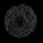
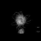
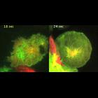
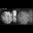
Fluorescence microscopy time series of Drosophila S2 cell stably expressing mCherry-alpha-tubulin and spreading on a ConA-coated substrate in the presence of cyto D. Long processes containing microtub...
CIL:40670
NCBI Organism Classification
Drosophila melanogaster
Biological Process
cytoskeleton organization
Cellular Component
microtubule
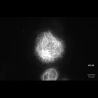
Time lapse movie prepared from fluorescence microscopy time series of a Drosophila S2 cell stably expressing mCherry-alpha-tubulin and spreading on a ConA-coated substrate in the presence of cyto D. L...
CIL:36797
NCBI Organism Classification
Drosophila melanogaster
Biological Process
microtubule cytoskeleton organization
Cellular Component
microtubule
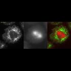
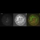
The first image in a z-series of a structured illumination image (SIM) of microtubules in a Drosophila S2 cell. The red channel is the diffraction limited image and the green channel is the SIM image...
CIL:11988
NCBI Organism Classification
Drosophila melanogaster
Biological Process
mitotic anaphase
Cellular Component
kinesin-6
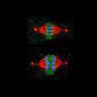
A comparison of kinesin-6 anaphase dynamics in a cell with a bipolar or monopolar spindle. (left) TIRF time lapse of kinesin-6 (Pav-GFP; green) and mCherry-tubulin (red) in a wild-type S2 cell with a ...
CIL:11984
NCBI Organism Classification
Drosophila melanogaster
Biological Process
mitotic anaphase
Cellular Component
myosin regulatory light chain
Time lapse of myosin GFP (left, imaged by TIRF microscopy) and mCherry tubulin (middle, imaged by epifluorescence; the spindle is close to the coverslip surface in this cell) during anaphase in a Dros...
CIL:191
NCBI Organism Classification
Drosophila melanogaster
Biological Process
mitosis
Cellular Component
spindle
The image shows mitotic metaphase (upper) and anaphase (lower) in Drosophila tissue culture cells immunostained for the microtubule severing protein katanin (green), microtubules (red) and kinetochore...
CIL:11992
NCBI Organism Classification
Drosophila melanogaster
Biological Process
mitotic anaphase
Cellular Component
myosin regulatory light chain
TIRF time lapse of mCherry-tubulin (left), myosin-GFP (middle), and overlay (myosin in green and tubulin in red; right) in a cell with a monopolar spindle from metaphase to anaphase. The S2 cells are...
CIL:11989
NCBI Organism Classification
Drosophila melanogaster
Biological Process
mitotic anaphase
Cellular Component
myosin regulatory light chain
TIRF time-lapse imaging of myosin-GFP (left) and mCherry-tubulin (right) in an S2 cell depleted of kinesin-6 (Pav) by RNAi. Myosin-GFP localization to the equator and loss from the poles does not occu...
« Previous
1
2
3
Next »
Results per page:
10
20
50
100