Licensing
Attribution Non-Commercial Share Alike:This image is licensed under a Creative Commons Attribution, Non-Commercial Share Alike License. View License Deed | View Legal Code
Comments:
Powered by Pnyxe
Description
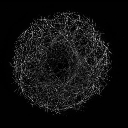
The first image in this multi-image tiff file is a structured illumination image (SIM) of microtubules in a Drosophila S2 cell. The second image is the corresponding diffraction limited image obtained with conventional fluorescence microscopy. Note the two-fold increase in spatial resolution in the SIM image compared to the diffraction limited image. The z-series are available as CIL 36797. Images were collected as part of the Marine Biological Laboratory Neurobiology Course, Summer 2011.
Technical Details
Cells were plated on Con-A coated coverslips, chemically fixed, and labeled with an anti-tubulin primary antibody and an Alexa Fluor 488 secondary antibody. Images were collected on a Zeiss Elyra Super-resolution microscope by Chris Rieken
Biological Sources
- NCBI Organism Classification
- Drosophila melanogaster
- (Common fruit fly)
- Cell Type
- epithelial cell
- Cell Line
- Schneider S2
- Cellular Component
- microtubule
Biological Context
- Biological Process
- microtubule cytoskeleton organization
Attribution
- Names
- Victor Cazares
- Luke Chao
- James A Galbraith
- Catherine G Galbraith
- Link
- MBL Neurobiology Course 2011
- Other
- MBL Neurobiology Course 2011
Grouping
This image is part of a group.Imaging
- Image Type
- recorded image
- Imaging Mode
- structured illumination microscopy (SIM)
- Parameters Imaged
- fluorescence emission
- Source of Contrast
- distribution of epitope
- Visualization Methods
- primary antibody plus labeled secondary antibody
- Alexa Fluor 488
- Processing History
- maximum projection
Sample Preparation
- Methods
- formaldehyde fixed tissue
- glutaraldehyde fixed tissue
- Relation To Intact Cell
- dispersed cells in vitro
Dimensions
| Spatial Axis | Image Size | Pixel Size |
|---|---|---|
| X | 1054px | 0.0397μm |
| Y | 1028px | 0.0397μm |
*CIL – Cell Image Library accession number. Please use this to reference an image.