Alternate header for print version
Advanced search
Contributors
Help
Submit
Search
menu
Cell Process
Cell Component
Cell Type
Organism
Microbial
Alzheimer's
Data Sets
University of California, San Diego
9500 Gilman Drive
La Jolla, CA 92093-0608, USA
Voice
: (858) 534-0276
Fax
: (858) 534-7497
Email
: dorloff@ncmir.ucsd.edu
Search Results for
establishment or maintenance of cel...
(356 results)
(Not the results you were expecting?)
(Comments)
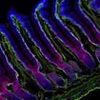
CIL:12291
NCBI Organism Classification
Mus musculus
Biological Process
cell-substrate adhesion
Cellular Component
microtubule
Colocalization of the microtubule anchoring factor LL5α (red) with integrin α6 (blue) in the basal cortex of outer epithelium of mouse mammary gland tissue. LL5 did not colocalize with β-catenin-p...
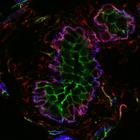
CIL:12293
NCBI Organism Classification
Mus musculus
Biological Process
cell-substrate adhesion
Cellular Component
microtubule
Colocalization of the microtubule anchoring factor LL5α (red) with integrin α6 (blue) and laminin (green) in the epithelium of mouse small intestine. LL5α is localized to the basal cell cortex atta...
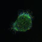
CIL:12294
NCBI Organism Classification
Homo sapiens
Biological Process
cell-substrate adhesion
Cellular Component
microtubule
Localization of the microtubule anchoring factor LL5α (red), microtubules (green), and the microtubule plus-end binding protein, EB1 (blue) in an MCF-10Aeco epithelial cell. EB1 was concentrated in ...
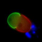
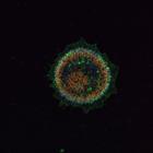
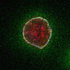
CIL:37933
NCBI Organism Classification
Caenorhabditis elegans
Biological Process
embryo development ending in birth or egg hatching
Cellular Component
cell cortex
Anterior and posterior regions of a C. elegans embryo are determined by PAR protein distribution. PAR-2, shown in green marks the posterior region of the embryo and PAR-6, shown in red, marks the ante...
CIL:12290
NCBI Organism Classification
Homo sapiens
Biological Process
cell-substrate adhesion
Cellular Component
microtubule
To determine the extent to which extracellular matrix proteins colocalize with the microtubule-anchoring factor LL5s (LL5α), MCF-10A epithelial cells were immunostained with antibodies to laminin rec...
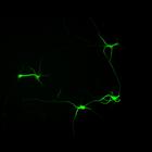
CIL:6162
NCBI Organism Classification
Rattus
Biological Process
neuron development
Cellular Component
cytoskeleton
Synapse formation in cultured hippocampal neurons after 7 days in vitro. Cells were immunostained for MAP2, a microtubule associated protein localized to dendrites but not axons (green), and synapsin...
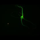
CIL:6187
NCBI Organism Classification
Rattus
Biological Process
neuron development
Cellular Component
cytoskeleton
Synapse formation in cultured hippocampal neurons after 7 days in vitro. Cells were immunostained for MAP2, a microtubule associated protein localized to dendrites but not axons (green), and synapsin...
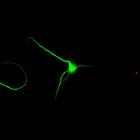
CIL:6199
NCBI Organism Classification
Rattus
Biological Process
neuron development
Cellular Component
cytoskeleton
Synapse formation in cultured hippocampal neurons after 7 days in vitro. Cells were immunostained for MAP2, a microtubule associated protein localized to dendrites but not axons (green), and synapsin...
CIL:12289
NCBI Organism Classification
Homo sapiens
Biological Process
cell-substrate adhesion
Cellular Component
microtubule
To determine the extent to which extracellular matrix proteins colocalize with the microtubule-anchoring factor LL5β, MCF-10A epithelial cells were immunostained with antibodies to the secreted autoc...
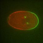
CIL:37932
NCBI Organism Classification
Caenorhabditis elegans
Biological Process
embryo development ending in birth or egg hatching
Cellular Component
cell cortex
Anterior and posterior regions of a C. elegans embryo are determined by PAR protein distribution. PAR-2, shown in green marks the posterior region of the embryo and PAR-6, shown in red, marks the ante...
« Previous
1
...
27
28
29
30
31
32
33
34
...
36
Next »
Results per page:
10
20
50
100