Licensing
Description
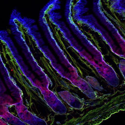
Colocalization of the microtubule anchoring factor LL5α (red) with integrin α6 (blue) and laminin (green) in the epithelium of mouse small intestine. LL5α is localized to the basal cell cortex attached to the laminin BMs. Overall findings from this publication showed that signaling from laminin-integrin associations play a role in attaching microtubule plus ends to the epithelial basal cell cortex. This image is the original data file from Fig. 4D, J Cell Biol (2010) 189 (5):901-917.
Technical Details
Small intestine from C57BL6/J strain mice (8 wk of age) was prepared by perfusion fixation, processed as described previously (Nakatani et al., 2004), and immunostained for laminin (Cy2, 488 nm laser), LL5a (rhodamine red-X, 543 nm laser) and integrin a6 (Cy5, 633 nm laser), and observed using a confocal laser-scanning microscope system.
Biological Sources
- NCBI Organism Classification
- Mus musculus
- (house mouse)
- Cell Type
- epithelial cell
- Cellular Component
- microtubule
- basal cortex
- microvillus
- basement membrane
Biological Context
Attribution
- Names
- Azusa Hotta
- Tomomi Kawakatsu
- Tomoya Nakatani
- Toshitaka Sato
- Chiyuki Matsui
- Taiko Sukezane
- Tsuyoshi Akagi
- Tomoki Hamaji
- Ilya Grigoriev
- Anna Akhmanova
- Yoshimi Takai
- Yuko Mimori-Kiyosue
- Published
- J. Cell Biol. 2010. 189(5):901-917
- Link
- http://jcb-dataviewer.rupress.org/jcb/...
- Pubmed
- 20513769
Grouping
This image is part of a group.Imaging
- Image Type
- recorded image
- Imaging Mode
- confocal microscopy
- Parameters Imaged
- fluorescence emission
- Source of Contrast
- distribution of epitope
- Visualization Methods
- Cy2
- X-Rhodamine
- Cy5
- Data Qualifiers
- raw, unprocessed data
- suitable for spatial measurements
Sample Preparation
- Methods
- formaldehyde fixed tissue
- Relation To Intact Cell
- microtome-sectioned tissue
Dimensions
| Spatial Axis | Image Size | Pixel Size |
|---|---|---|
| X | 1024px | 0.45μm |
| Y | 1024px | 0.45μm |