Alternate header for print version
Advanced search
Contributors
Help
Submit
Search
menu
Cell Process
Cell Component
Cell Type
Organism
Microbial
Alzheimer's
Data Sets
University of California, San Diego
9500 Gilman Drive
La Jolla, CA 92093-0608, USA
Voice
: (858) 534-0276
Fax
: (858) 534-7497
Email
: dorloff@ncmir.ucsd.edu
Search Results for
columnar/cuboidal epithelial cell
(1212 results)
(Not the results you were expecting?)
(Comments)
CIL:36076
NCBI Organism Classification
Felis catus
Biological Process
intestinal absorption
Cellular Component
microvillus
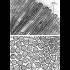
Figures 446 (upper) and 447 (lower) from Chapter 16 (Cytoplasmic matrix and cytoskeleton) of 'The Cell, 2nd Ed.' by Don W. Fawcett M.D. Microvilli of the brush border of cat intestinal epithelium sho...
CIL:11106
NCBI Organism Classification
Rattus
Biological Process
cell projection organization
Cellular Component
brush border
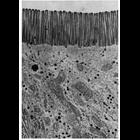
Electron micrograph showing the brush border of intestinal epithelial cells of the rat. The parallel microvilli of the brush border are about 0.1 µm wide and 1.0 µm in length. Image by Jean Paul R...
CIL:41544
NCBI Organism Classification
Homo sapiens
Biological Process
none specified
Cellular Component
hyaline layer
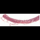
Histology sample illustrating the pseudostratified, ciliated columnar epithelium of the trachea. Note that the basement membrane underlying this particular epithelium is especially prominent. This im...
CIL:11534
NCBI Organism Classification
Macaca mulatta
Biological Process
pigmentation
Cellular Component
melanosome
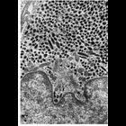
Figure 299 from Chapter 11 (Melanin Pigment) of 'The Cell, 2nd Ed.' by Don W. Fawcett M.D. Melanocyte pigment cell from the iris in the eye of the Macaca mulatta. The cytoplasm is crowded with melan...
CIL:38956
NCBI Organism Classification
none specified
Biological Process
melanosome localization
Cellular Component
melanosome
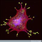
Confocal micrograph of an isolated melanin-producing cell (a melanocyte) showing the melanosomes (vesicles that hold the melanin granules) in yellow, the actin in red and the microtubules in blue. The...
CIL:38978
NCBI Organism Classification
Homo sapiens
Biological Process
mitosis
Cellular Component
endoplasmic reticulum
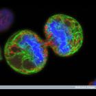
Human melanoma cell undergoing cell division. The chromosomes (blue) have separated and the two daughter cells have almost split apart - only a small bridge of cytoplasm remains. The green staining la...
CIL:10944
NCBI Organism Classification
Homo sapiens
Biological Process
cell-substrate adhesion
Cellular Component
basal lamina
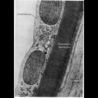
This electron micrograph shows the endothelium and the underlying Descemet's membrane of the human cornea. Descemet's membrane is an unusually thick and structurally specialized example of basal lami...
CIL:25776
NCBI Organism Classification
Platynereis dumerilii
Biological Process
convergent extension involved in axis elongation
Cellular Component
none specified
Polarized cell rearrangement in the Platynereis neuroectoderm visualized by time-lapse imaging. This time-lapse movie shows convergent extension by mediolateral cell intercalation of the ventral neuro...
CIL:10931
NCBI Organism Classification
Myotis
Biological Process
extracellular structure organization
Cellular Component
plasma membrane
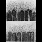
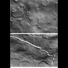
Filaments extend from microvilli of the epithelial cells in the intestine of the bat, Moytis lucifugus in these two high magnification electron micrographs. These filaments, which are 2.5-5nm thick, ...
CIL:11237
NCBI Organism Classification
Macaca mulatta
Biological Process
cell communication
Cellular Component
gap junction
Representative examples of gap junctions from vertebrates (ciliary epithelium from the eye of a Macaca mulatta, upper) and invertebrates (inverted gap junctions from cells of the mid-gut of the horses...
« Previous
1
2
3
4
5
6
7
8
9
...
122
Next »
Results per page:
10
20
50
100