Licensing
Description
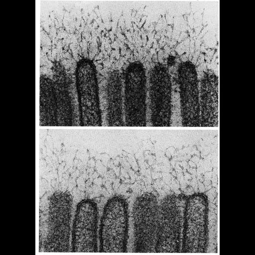
Filaments extend from microvilli of the epithelial cells in the intestine of the bat, Moytis lucifugus in these two high magnification electron micrographs. These filaments, which are 2.5-5nm thick, and extend from 0.1 - 0.5µm beyond the microvilli, make up the glycocalyx that encompasses the brush border of the intestine. Filaments from each microvilli radiate and intermingle with those from adjacent microvilli to form a near-continuous network. Micrographs courtesy of Susumu Ito; upper panel was reprinted with permission from Fawcett, D. J. (1965) Histochem. Cytochem. 13: 75-95, and are figures 17 (upper) and 18 (lower) from Chapter 1 (The Cell Surface) of 'The Cell, 2nd Ed.' by Don W. Fawcett M.D. A PDF copy of the accompanying chapter is available on the ASCB's BioEDUCATE website.
Biological Sources
- NCBI Organism Classification
- Myotis
- (mouse-eared bats)
- Cell Type
- epithelial cell
- enterocyte
- Cellular Component
- plasma membrane
- extracellular region part
- glycocalyx
- microvillus
Biological Context
- Biological Process
- extracellular structure organization
Attribution
- Names
- Don W. Fawcett
- Susumu Ito
- Published
- ISBN 0721635849
- Links
- The Cell, 2nd Ed. by D.W. Fawcett
- J Histochem Cytochem reprint
- Pubmed
- 14293043
Grouping
This image is part of a group.Imaging
- Image Type
- transmission electron microscopy (TEM)
- Imaging Mode
- illumination by electrons
- detection of electrons
- Parameters Imaged
- electron density
- Source of Contrast
- stain with broad specificity
- Data Qualifiers
- processed data
Sample Preparation
- Methods
- crosslinking-fixative fixed tissue
- Relation To Intact Cell
- microtome-sectioned tissue
Dimensions
| Spatial Axis | Image Size | Pixel Size |
|---|---|---|
| X | 918px | —— |
| Y | 1268px | —— |