Alternate header for print version


Licensing
Attribution Non-Commercial Share Alike:This image is licensed under a Creative Commons Attribution, Non-Commercial Share Alike License. View License Deed | View Legal Code
tweet  
*CIL – Cell Image Library accession number. Please use this to reference an image.

CIL:41544*  Cite 
Description

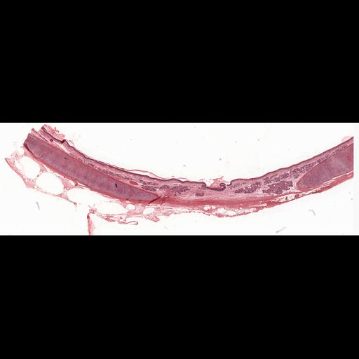
Histology sample illustrating the pseudostratified, ciliated columnar epithelium of the trachea. Note that the basement membrane underlying this particular epithelium is especially prominent. This image is slide 40 in the University of Michigan Histology and Virtual Learning Resource. A more detailed description can be found by following the indicated url link http://histology.med.umich.edu/medical/connective-tissue.

Technical Details

Biological Sources
NCBI Organism Classification
Homo sapiens
Cell Type
ciliated epithelial cell
Cellular Component
hyaline layer
Citation
Digital Object Identifier (DOI)
doi:10.7295/W9CIL41544
Archival Resource Key (ARK)
ark:/b7295/w9cil41544
Imaging
Image Type
recorded image
Image Mode
bright-field microscopy
Parameters Imaged
absorption of illumination
Source of Contrast
differences in adsorption or binding of stain
Visualization Methods
hematoxylin
eosin
Sample Preparation
Methods
chemically fixed tissue
Relation To Intact Cell
sectioned tissue
Dimensions
Spatial Axis Image Size Pixel Size
X 4000px ——
Y 1264px ——