Alternate header for print version
Advanced search
Contributors
Help
Submit
Search
menu
Cell Process
Cell Component
Cell Type
Organism
Microbial
Alzheimer's
Data Sets
Center for Research in Biological Systems
University of California, San Diego
9500 Gilman Drive
La Jolla, CA 92093-0608, USA
Voice
: (858) 534-0276
Fax
: (858) 534-7497
Email
: dorloff@ncmir.ucsd.edu
Search Results for
"plasma membrane"
(1457 results)
(Not the results you were expecting?)
(Comments)
Still Images
Video/Animation
Z-Stack
Time Series
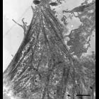
CIL:11237
NCBI Organism Classification
Macaca mulatta
Biological Process
cell communication
Cellular Component
gap junction
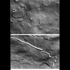
Representative examples of gap junctions from vertebrates (ciliary epithelium from the eye of a Macaca mulatta, upper) and invertebrates (inverted gap junctions from cells of the mid-gut of the horses...
CIL:11211
NCBI Organism Classification
Opsanus tau
Biological Process
cell adhesion
Cellular Component
desmosome
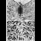
Upper: capillary endothelial cell junction in the rete mirabile of the gas bladder of the toadfish Opsanus tau shows dense intracelluular plaques and the central dense line in the intercellular space,...
CIL:12103
NCBI Organism Classification
Colpoda cucullus
Biological Process
cell motility
Cellular Component
cilium
A layer of mucocysts is docked under the pellicle. The pellicle has a plasma membrane and an alveolar sac system. Both basal bodies of a dikinetid are ciliated, and the food vacuoles are plentiful and...
CIL:12633
NCBI Organism Classification
Paramecium multimicronucleatum
Biological Process
early endosome to Golgi transport
Cellular Component
clathrin coat of endocytic vesicle
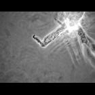
A high resolution of a quick-freeze deep-etch rotary shadowed replica of a parasomal sac from which a clathrin-coated preendosomal vesicle has pinched off. The typical clathrin cage structure is prese...
CIL:12599
NCBI Organism Classification
Canis lupus familiaris
Biological Process
none specified
Cellular Component
stress fiber
Network of stress fibers in an MDCK cell. This transmission electron micrograph of a flat-embedded MDCK cell was taken 60-70nm from the interface between the cell and the coverslip on which it was gro...
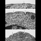
CIL:13003
NCBI Organism Classification
Cavia porcellus
Biological Process
pinocytosis
Cellular Component
coated vesicle
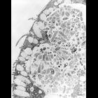
Stages of coated pit vesicle invagination during micropinocytosis. Each frame is a portion of the surface of polychromatophilic erythroblasts from guinea pig bone marrow and various stages of vesicle...
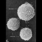
CIL:13091
NCBI Organism Classification
Mus musculus
Biological Process
immune system process
Cellular Component
microvillus
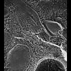
This image shows two different types of white blood cells from a mouse which are essential for mammalian immune response to protect from infection. The larger cell is a macrophage and the smaller two...
CIL:13698
NCBI Organism Classification
Chlorocebus aethiops
Biological Process
G-protein coupled receptor internalization
Cellular Component
plasma membrane
This is one of a group of four images in Figure S5 in Apaja et al., JCB 2010, that support the conclusion that the WT vasopressin type2 receptor (V2R) and the V2R W164S mutant (that causes nephrogenic...
CIL:24574
NCBI Organism Classification
Balamuthia mandrillaris
Biological Process
pseudopodium organization
Cellular Component
cell
A frame from the grouped movie of a Balamuthia mandrillaris ameba within a culture of monkey kidney cells. Accompanying photographs are enlargements of the tips of pseudopodia to demonstrate their fle...
CIL:258
NCBI Organism Classification
Mus musculus
Biological Process
plasma membrane organization
Cellular Component
dystroglycan complex
Thin section electron microscopy of diaphragm (skeletal) muscle from a wild type mouse. Sample was viewed with a Hitachi 7600 electron microscope (accelerating voltage 80 KV) and imaged with an AMT d...
« Previous
1
...
38
39
40
41
42
43
44
45
...
146
Next »
Results per page:
10
20
50
100