Alternate header for print version
Advanced search
Contributors
Help
Submit
Search
menu
Cell Process
Cell Component
Cell Type
Organism
Microbial
Alzheimer's
Data Sets
University of California, San Diego
9500 Gilman Drive
La Jolla, CA 92093-0608, USA
Voice
: (858) 534-0276
Fax
: (858) 534-7497
Email
: dorloff@ncmir.ucsd.edu
Search Results for
optical path length gradient
(1067 results)
(Not the results you were expecting?)
(Comments)
CIL:24801
NCBI Organism Classification
Xenopus laevis
Biological Process
cellular localization
Cellular Component
lamellipodium
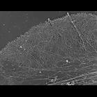
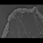
Structural differentiation of actin network in lamellipodium. Electron micrograph of Xenopus fibroblasts after regular extraction in the presence of polyethelene glycol (PEG) and phalloidin. While the...
CIL:24802
NCBI Organism Classification
Xenopus laevis
Biological Process
cellular localization
Cellular Component
lamellipodium
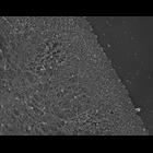
Structural differentiation of actin network in lamellipodium. Electon micrograph Xenopus fibroblast after unprotected extraction without polyethelene glycol. Actin network at lamellipodial rear disas...
CIL:24808
NCBI Organism Classification
Xenopus laevis
Biological Process
cellular localization
Cellular Component
lamellipodium
Localization of XAC (Xenopus ADF/cofilin) in Xenopus keratocytes done with immuno-EM. A low mag view of the cell from which this high mag view is taken is shown in CIL 24807. Image corresponds to Fi...
CIL:24809
NCBI Organism Classification
Xenopus laevis
Biological Process
cellular localization
Cellular Component
lamellipodium
Localization of Xenopus ADF/cofilin (XAC) to posterior regions of depolymerization-resistant actin brush. Electron (micrograph of lamellipodia of Xenopus keratocytes after unprotected extraction and s...
CIL:24811
NCBI Organism Classification
Xenopus laevis
Biological Process
cellular localization
Cellular Component
lamellipodium
Localization of Xenopus ADF/cofilin (XAC) to posterior regions of depolymerization-resistant actin brush. Electron micrograph of lamellipodia of Xenopus keratocytes after latrunculin a treatment ( 0....
CIL:24790
NCBI Organism Classification
none specified
Biological Process
branching of actin filaments
Cellular Component
lamellipodium
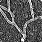
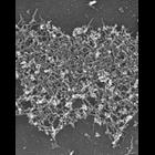
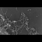
Improved visualization of actin filament branching in lamellipodia. EM of keratocyte or fibroblast lamellipodial actin network after cytochalasin D treatment (0.2 μM for 30 min or 0.5 μM for 10 min)...
CIL:27219
NCBI Organism Classification
Homo sapiens
Biological Process
none specified
Cellular Component
cell
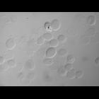
This image set consists of five differential interference contrast (DIC) images of red bood cells (approximately 8 microns in diameter). Each image in the set contains a DIC image and a thresholded i...
CIL:35065
NCBI Organism Classification
none specified
Biological Process
branching of actin filaments
Cellular Component
actin cytoskeleton
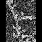
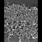
Electron micrograph of keratocyte or fibroblast lamellipodial actin network after unprotected extraction. All examples demonstrate frequent branching of actin filaments. Image corresponds to a singl...
CIL:35066
NCBI Organism Classification
none specified
Biological Process
branching of actin filaments
Cellular Component
actin cytoskeleton
Electron micrograph of keratocyte or fibroblast lamellipodial actin network after unprotected extraction. All examples demonstrate frequent branching of actin filaments. Image corresponds to a singl...
CIL:34895
NCBI Organism Classification
none specified
Biological Process
branching of actin filaments
Cellular Component
actin cytoskeleton
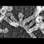
Improved visualization of actin filament branching in lamellipodia. EM of keratocyte or fibroblast lamellipodial actin network after cytochalasin D treatment (0.2 μM for 30 min or 0.5 μM for 10 min)...
« Previous
1
...
9
10
11
12
13
14
15
16
...
107
Next »
Results per page:
10
20
50
100

")