Licensing

Attribution_nc_sa
Attribution Non-Commercial Share Alike:This image is licensed under a Creative Commons Attribution, Non-Commercial Share Alike License. View License Deed | View Legal Code
Comments: Powered by Pnyxe

Description

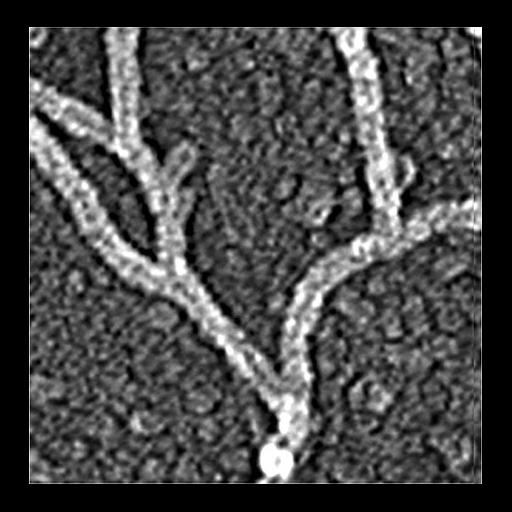
Electron micrograph of keratocyte or fibroblast lamellipodial actin network after unprotected extraction. All examples demonstrate frequent branching of actin filaments. Image corresponds to a single panel from Figure 2d in J Cell Biol. 1999 May 31;145(5):1009-26. All of the panels from Figure 2d can be found as CIL 35060-35068.

Technical Details

Procedures for detergent extraction, immunostaining, S1 decoration, light, and EM were described previously (Svitkina et al., 1995, 1996, 1997;Verkhovsky et al., 1995; Svitkina and Borisy, 1998).

Biological Sources

Cellular Component
actin cytoskeleton

Biological Context

Biological Process
branching of actin filaments
actin filament-based process
Molecular Function
structural constituent of cytoskeleton

Attribution

Names
Tatyana M. Svitkina
Gary G. Borisy
Published
J Cell Biol. 1999 May 31;145(5):1009-26.
Pubmed
10352018

Grouping

This image is part of a group.

Imaging

Image Type
recorded image
Imaging Mode
transmission electron microscopy (TEM)
Parameters Imaged
optical path length gradient
Source of Contrast
boundaries between regions with different refractive index
Visualization Methods
shadowing and plating
platinum replica
Processing History
unprocessed raw data

Sample Preparation

Methods
permeabilized tissue
glutaraldehyde fixed tissue
Relation To Intact Cell
dispersed cells in vitro

Dimensions

Spatial Axis Image Size Pixel Size
X 453px ——
Y 457px ——
*CIL – Cell Image Library accession number. Please use this to reference an image.