Alternate header for print version
Advanced search
Contributors
Help
Submit
Search
menu
Cell Process
Cell Component
Cell Type
Organism
Microbial
Alzheimer's
Data Sets
University of California, San Diego
9500 Gilman Drive
La Jolla, CA 92093-0608, USA
Voice
: (858) 534-0276
Fax
: (858) 534-7497
Email
: dorloff@ncmir.ucsd.edu
Search Results for
confocal microscopy
(2394 results)
(Not the results you were expecting?)
(Comments)
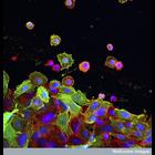
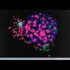
CIL:38917
NCBI Organism Classification
none specified
Biological Process
cell migration
Cellular Component
actin cytoskeleton
Confocal micrograph of cultured colon cancer cells showing the nuclei stained with DAPI in blue, the actin cytoskeleton in red and plectin (isoform 1k) in green. Plectin interacts with cytoskeletal ac...
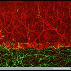
CIL:38933
NCBI Organism Classification
none specified
Biological Process
cerebellar Purkinje cell layer morphogenesis
Cellular Component
neuron projection
This confocal micrograph shows specialized cells named Purkinje cells (red) that are found in a part of the brain called the cerebellum. They send out vast numbers of branches that make connections wi...
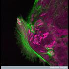
CIL:38936
NCBI Organism Classification
Shigella
Biological Process
actin polymerization-dependent cell motility
Cellular Component
actin cytoskeleton
Confocal micrograph showing Shigella bacteria (pink) invading the intestinal lining. The bacteria infects the cells by high-jacking the cell's internal actin skeleton (green) to facilitate its entry i...
CIL:38939
NCBI Organism Classification
Mumps virus
Biological Process
response to virus
Cellular Component
endoplasmic reticulum
This confocal micrograph shows the mumps virus protein (turquoise) in the endoplasmic reticulum of a cultured cell. This is a region of the cell that processes proteins. This particular protein is pos...
CIL:39007
NCBI Organism Classification
Homo sapiens
Biological Process
fertilization
Cellular Component
gap junction
A confocal micrograph of a human embryo, with some remaining sperm cells, about five days after fertilization. The nuclei of both the sperm and cells of the embryo are stained purple, the tails of the...
CIL:39058
NCBI Organism Classification
none specified
Biological Process
neuron migration
Cellular Component
actin cytoskeleton
This confocal micrograph shows a dorsal root ganglion (DRG) explant. The dorsal root ganglion is a swelling on the dorsal roots of spinal nerves, which contains a cluster of cell bodies and synapses. ...
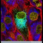
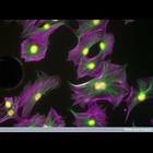
CIL:39060
NCBI Organism Classification
none specified
Biological Process
microtubule cytoskeleton organization
Cellular Component
microtubule
Confocal micrograph of osteoblast cells labeled with Alexafluor 488 that stains alpha tubulin (green) and phalloidin marking the actin (purple) and DAPI highlighting the nucleus (yellow). Osteoblasts...
CIL:39023
NCBI Organism Classification
Danio rerio
Biological Process
reticulospinal neuron morphogenesis
Cellular Component
neuronal cell body
Confocal micrograph of a transverse section of the brain of a zebrafish embryo, at the level of the hindbrain. The brain section was stained using an antibody that marks a calcium binding protein (cal...
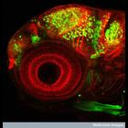
CIL:39027
NCBI Organism Classification
Danio rerio
Biological Process
Dlx4/6 promotor specification
Cellular Component
neuron projection
Confocal micrograph of the head region of a transgenic zebrafish embryo. The large circular structure is the eye, showing the structure of the retina. Some neurons in the brain are highlighted in gre...
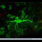
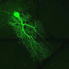
CIL:40124
NCBI Organism Classification
Mus musculus
Biological Process
cerebellum structural organization
Cellular Component
cell projection cytoplasm
Purkinje neuron from mouse cerebellum injected with Lucifer Yellow and imaged using confocal microscopy. This image represents a mosaic of 4 different volumes, and has been downsampled from the raw da...
« Previous
1
...
17
18
19
20
21
22
23
24
...
240
Next »
Results per page:
10
20
50
100