Licensing

Attribution_by_nc_nd
Attribution Non-Commercial; No Derivatives:This image is licensed under a Creative Commons Attribution, Non-Commercial, No Derivatives License. View License Deed | View Legal Code
Comments: Powered by Pnyxe

Description

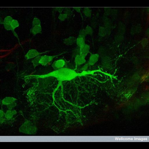
Confocal micrograph of a transverse section of the brain of a zebrafish embryo, at the level of the hindbrain. The brain section was stained using an antibody that marks a calcium binding protein (calretinin). The image shows a big reticulospinal neuron and its processes, surrounded by other smaller neurons. Reticulospinal neurons are specific neurons found in the brains of fish that are thought to be involved in coordinating swimming.

Technical Details

B0007775 Reticulospinal neuron. Wellcome Images available under the following creative commons usage http://creativecommons.org/licenses/by-nc-nd/2.0/uk/

Biological Sources

NCBI Organism Classification
Danio rerio
(zebrafish)
Cell Type
reticulospinal neuron
Cellular Component
neuronal cell body
neuron projection
calretinin

Biological Context

Biological Process
reticulospinal neuron morphogenesis
Zebrafish Anatomy
hindbrain

Attribution

Name
Steve Wilson
Link
Wellcome Images

Imaging

Image Type
recorded image
Imaging Mode
confocal microscopy
Parameters Imaged
fluorescence emission
Source of Contrast
distribution of epitope
Visualization Methods
primary antibody plus labeled secondary antibody
Processing History
unprocessed raw data

Sample Preparation

Relation To Intact Cell
sectioned tissue

Dimensions

Spatial Axis Image Size Pixel Size
X 635px ——
Y 576px ——
*CIL – Cell Image Library accession number. Please use this to reference an image.