Alternate header for print version


Licensing
Attribution Non-Commercial; No Derivatives:This image is licensed under a Creative Commons Attribution, Non-Commercial, No Derivatives License. View License Deed | View Legal Code
tweet  
*CIL – Cell Image Library accession number. Please use this to reference an image.

CIL:38936*  Cite 
Description

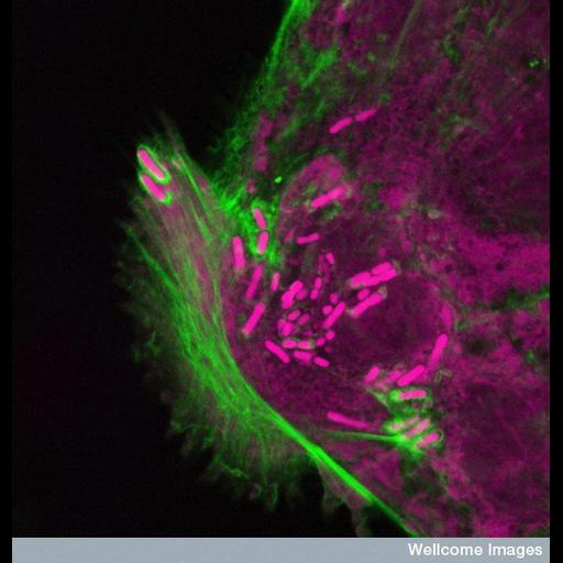
Confocal micrograph showing Shigella bacteria (pink) invading the intestinal lining. The bacteria infects the cells by high-jacking the cell's internal actin skeleton (green) to facilitate its entry into the cell and spread into adjoining cells, using polymerizing actin comet tails as several can be seen doing here. Shigella intestinal infection causes diarrhea and rapid dehydration typical of bacterial dysentery. FITC-labelled phalloidin highlights the actin of the cytoskeleton in green and propidium iodide (a DNA stain) was used to visualise bacteria in pink.

Technical Details

B0006238 Shigella infection. Wellcome Images available under the following creative commons usage http://creativecommons.org/licenses/by-nc-nd/2.0/uk/

Biological Sources
NCBI Organism Classification
Shigella
Homo sapiens
Cell Type
intestinal epithelial cell
Cellular Component
actin cytoskeleton
DNA
comet tails
Biological Context
Biological Process
actin polymerization-dependent cell motility
Molecular Function
bacterial invasion
Human Disease
shigellosis
diarrhea
Attribution
Names
S. Schuller
Link
Wellcome Images
Citation
Digital Object Identifier (DOI)
doi:10.7295/W9CIL38936
Archival Resource Key (ARK)
ark:/b7295/w9cil38936
Imaging
Image Type
recorded image
Image Mode
confocal microscopy
Parameters Imaged
fluorescence microscopy
Source of Contrast
distribution of epitope
Visualization Methods
propidium iodide
phalloidin
Fluorescein (FITC)
Sample Preparation
Methods
chemically fixed tissue
Dimensions
Spatial Axis Image Size Pixel Size
X 550px ——
Y 576px ——