Alternate header for print version
Advanced search
Contributors
Help
Submit
Search
menu
Cell Process
Cell Component
Cell Type
Organism
Microbial
Alzheimer's
Data Sets
University of California, San Diego
9500 Gilman Drive
La Jolla, CA 92093-0608, USA
Voice
: (858) 534-0276
Fax
: (858) 534-7497
Email
: dorloff@ncmir.ucsd.edu
Search Results for
dendrite development
(361 results)
(Not the results you were expecting?)
(Comments)
CIL:40659
NCBI Organism Classification
Rattus
Biological Process
dendritic spine organization
Cellular Component
dendritic spine
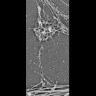
Platinum replica depicting the cytoskeletal organization of dendritic spines in extracted 14 DIV neurons. Mushroom spines associate with dendrites at the base (bottom) and with axons by the head (top)...
CIL:40661
NCBI Organism Classification
Rattus
Biological Process
dendritic spine organization
Cellular Component
dendritic spine
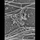
Platinum replica depicting the cytoskeletal organization of dendritic spines in extracted 14 DIV neurons. This image shows the interaction of putative PSD from the spine head with axonal intermediate ...
CIL:40804
NCBI Organism Classification
Rattus
Biological Process
dendrite morphogenesis
Cellular Component
axon
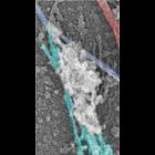
Colorized transmission electron micrograph of a platinum replica showing the cytoskeletal organization of stubby dendritic spines in extracted hippocampal neurons after 14 DIV. This image shows axons,...
CIL:40662
NCBI Organism Classification
Rattus
Biological Process
dendritic spine organization
Cellular Component
dendritic spine
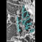
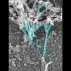
Platinum replica depicting the cytoskeletal organization of dendritic spines in extracted 14 DIV neurons. Branched actin filaments (cyan) in the head. Image corresponds to Figure 2a in Mol Biol Cell....
CIL:40663
NCBI Organism Classification
Rattus
Biological Process
dendritic spine organization
Cellular Component
dendritic spine
Platinum replica depicting the cytoskeletal organization of dendritic spines in extracted 14 DIV neurons. This image shows branched actin filaments (cyan) at the neck–head junction. Dashed arrows ...
CIL:10270
NCBI Organism Classification
Rattus
Biological Process
cellular developmental process
Cellular Component
dendrite
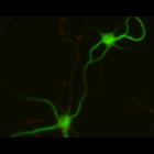
Early stages of dendritic development and synapse formation in cultured hippocampal neurons. This multilayer image shows neurons fixed at 7 days in vitro and immunostained for the dendritically loca...
CIL:10316
NCBI Organism Classification
Rattus
Biological Process
cellular developmental process
Cellular Component
dendrite
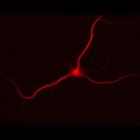
Cultured hippocampal neurons after 10 days in vitro, immunostained for MAP2, a microtubule associated protein localized to dendrites (red) but not axons, which are not apparent in the immunofluorescen...
CIL:10349
NCBI Organism Classification
Rattus
Biological Process
cellular developmental process
Cellular Component
dendrite
Cultured hippocampal neurons after 14 days in vitro, immunostained for MAP2, a microtubule associated protein localized to dendrites (red) but not axons, which are not apparent in the immunofluorescen...
CIL:10113
NCBI Organism Classification
Rattus
Biological Process
developmental process
Cellular Component
cytoskeleton
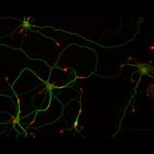
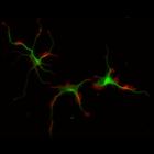
This multi-layer image shows the spatial relationship between filamentous actin (red) and microtubule array (green) in cultured hippocampal neurons, grown for 5 days in vitro. Actin staining (with rh...
CIL:10208
NCBI Organism Classification
Rattus
Biological Process
developmental process
Cellular Component
cytoskeleton
This multi-layer image shows the spatial relationship between filamentous actin (red) and microtubule array (green) in cultured hippocampal neurons, grown for 1 day in vitro. Actin staining (with rho...
« Previous
1
...
10
11
12
13
14
15
16
17
...
37
Next »
Results per page:
10
20
50
100