Licensing
Attribution Non-Commercial Share Alike:This image is licensed under a Creative Commons Attribution, Non-Commercial Share Alike License. View License Deed | View Legal Code
Comments:
Powered by Pnyxe
Description
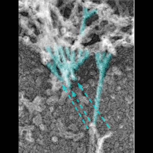
Platinum replica depicting the cytoskeletal organization of dendritic spines in extracted 14 DIV neurons. This image shows branched actin filaments (cyan) at the neck–head junction. Dashed arrows indicate potential filament breakage. Image corresponds to Figure 2a in Mol Biol Cell. 2010 Jan 1;21(1):165-76. The entire panel is available as CIL 40660. The uncolorized image is available as CIL 40659. Box 1, 2, and 3 (this image) are CIL 40661, 40662, and 40663, respectively.
Technical Details
Please refer to referenced article for details on cell extraction and processing for electron microscopy.
Biological Sources
- NCBI Organism Classification
- Rattus
- (Old World rats)
- Cell Type
- hippocampal neuron
- Cellular Component
- dendritic spine
- dendritic shaft
- actin cytoskeleton
Biological Context
- Biological Process
- dendritic spine organization
- dendrite morphogenesis
Attribution
- Names
- Farida Korobova
- Tatyana Svitkina
- Published
- Mol Biol Cell. 2010 Jan 1;21(1):165-76. Epub 2009 Nov 4.
- Pubmed
- 19889835
Grouping
This image is part of a group.Imaging
- Image Type
- grey scale graphic
- Imaging Mode
- transmission electron microscopy (TEM)
- Parameters Imaged
- electron density
- Source of Contrast
- differences in deposition of metal shadow
- Visualization Methods
- shadowing and plating
- Processing History
- colorized
Sample Preparation
- Methods
- detergent permeabilized
- glutaraldehyde fixed tissue
- critical_point dried specimen
- Relation To Intact Cell
- dispersed cells in vitro
Dimensions
| Spatial Axis | Image Size | Pixel Size |
|---|---|---|
| X | 464px | —— |
| Y | 612px | —— |
*CIL – Cell Image Library accession number. Please use this to reference an image.