Alternate header for print version


Licensing
Attribution Non-Commercial Share Alike:This image is licensed under a Creative Commons Attribution, Non-Commercial Share Alike License. View License Deed | View Legal Code
tweet  
*CIL – Cell Image Library accession number. Please use this to reference an image.

CIL:40659*  Cite 
Description

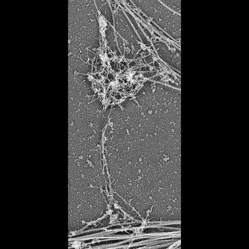
Platinum replica depicting the cytoskeletal organization of dendritic spines in extracted 14 DIV neurons. Mushroom spines associate with dendrites at the base (bottom) and with axons by the head (top). Thick fibers in neurites represent microtubules. Image corresponds to Figure 2a in Mol Biol Cell. 2010 Jan 1;21(1):165-76. The entire panel is available as CIL 40660. The uncolorized image (this image) is available as CIL 40659. Box 1, 2, and 3 are CIL 40661, 40662, and 40663, respectively.

Technical Details

Please refer to referenced article for details on cell extraction and processing for electron microscopy.

Biological Sources
NCBI Organism Classification
Rattus
Cell Type
hippocampal neuron
Cellular Component
dendritic spine
dendritic shaft
post synaptic density
actin cytoskeleton
microtubule cytoskeleton
intermediate filament cytoskeleton
Biological Context
Biological Process
dendritic spine organization
dendrite morphogenesis
Attribution
Names
Farida Korobova
Tatyana Svitkina
Published
Mol Biol Cell. 2010 Jan 1;21(1):165-76. Epub 2009 Nov 4.
Pubmed
19889835
Citation
Digital Object Identifier (DOI)
doi:10.7295/W9CIL40659
Archival Resource Key (ARK)
ark:/b7295/w9cil40659
Grouping This image is part of a group.
Imaging
Image Type
grey scale graphic
Image Mode
transmission electron microscopy (TEM)
Parameters Imaged
electron density
Source of Contrast
differences in deposition of metal shadow
Visualization Methods
shadowing and plating
Processing History
unprocessed raw data
Dimensions
Spatial Axis Image Size Pixel Size
X 891px ——
Y 1965px ——