Alternate header for print version
Advanced search
Contributors
Help
Submit
Search
menu
Cell Process
Cell Component
Cell Type
Organism
Microbial
Alzheimer's
Data Sets
University of California, San Diego
9500 Gilman Drive
La Jolla, CA 92093-0608, USA
Voice
: (858) 534-0276
Fax
: (858) 534-7497
Email
: dorloff@ncmir.ucsd.edu
Search Results for
Drosophila melanogaster
(437 results)
(Not the results you were expecting?)
(Comments)
CIL:32144
NCBI Organism Classification
Drosophila melanogaster
Biological Process
cellular localization
Cellular Component
nucleus
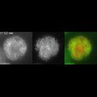
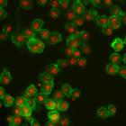
Drosophila melanogaster Kc167 cells were stained for DNA (to label nuclei, red) and actin (a cytoskeletal protein, to show the cell body, green). Each image is a dual channel fluorescent image followe...
CIL:25607
NCBI Organism Classification
Drosophila melanogaster
Biological Process
innate immune response
Cellular Component
none specified
Loose tethering of blood cells adherent to the unwounded body wall of a Drosophila third-instar larva. Real-time movie of blood cells seen from the dorsal side of a Nrg-GFP;PxnYFP larva. A cluster of ...
CIL:25610
NCBI Organism Classification
Drosophila melanogaster
Biological Process
innate immune response
Cellular Component
none specified
Blood-cell flow in dorsal region of a Drosophia third-instar larval body. Live visualization of blood cell flow in a Nrg-GFP;PxnYFP larva viewed dorsally. Most blood cells flow from anterior (Upper) t...
CIL:11987
NCBI Organism Classification
Drosophila melanogaster
Biological Process
mitotic anaphase
Cellular Component
myosin regulatory light chain
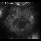
TIRF time lapse of myosin-GFP (left), mCherry-tubulin (middle), and overlay (myosin-GFP in green and mCherry-tubulin in red) in wild-type S2 cells from metaphase to anaphase. This cell has many astral...
CIL:11993
NCBI Organism Classification
Drosophila melanogaster
Biological Process
mitotic anaphase
Cellular Component
myosin regulatory light chain
TIRF time lapse of myosin-GFP in a cell with a symmetric monopolar spindle from metaphase to anaphase (cell was depleted of Klp61F [kinesin-5] and BubR1 by RNAi). At anaphase, myosin-GFP concentrates ...
CIL:13469
NCBI Organism Classification
Drosophila melanogaster
Biological Process
endocytosis
Cellular Component
early endosome
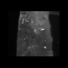
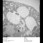
Transmission electron micrograph of Drosophila nephrocyte garland cell from ema[1] mutant: high magnification view. ema mutant garland cells are filled with very large electron-lucent alpha and electr...
CIL:13475
NCBI Organism Classification
Drosophila melanogaster
Biological Process
receptor-mediated endocytosis
Cellular Component
endosome
The endosomal compartment is enlarged in the ema[1] mutant. Colocalization between DVGLUT (red) aggregates and the endosomal ESCRT protein Hrs (green, also known as Vps27) in motoneuron cell bodies of...
CIL:13477
NCBI Organism Classification
Drosophila melanogaster
Biological Process
endocytosis
Cellular Component
endosome
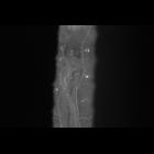
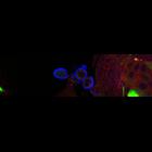
Ema localizes to endosomes. Third instar larval garland cells expressing Ema-GFP fusion protein and stained for the late endosomal and lysosomal protein Spinster (red) and plasma membrane and nuclei (...
CIL:13478
NCBI Organism Classification
Drosophila melanogaster
Biological Process
endocytosis
Cellular Component
endosome
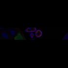
Ema localizes to endosomes. Third instar larval garland cells expressing Ema-GFP fusion protein and stained for the early endosomal protein Rab5 (red) and plasma membrane and nuclei (blue). Ema does n...
CIL:13676
NCBI Organism Classification
Drosophila melanogaster
Biological Process
response to wounding
Cellular Component
nucleus
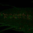
The JNK phosphatase puckered, whose expression can be reported via a lacZ enhancer trap, is expressed in very low levels in uninjured neurons; however, nerve crush induces a dramatic increase in puc-l...
« Previous
1
...
35
36
37
38
39
40
41
42
...
44
Next »
Results per page:
10
20
50
100
")