Alternate header for print version
Advanced search
Contributors
Help
Submit
Search
menu
Cell Process
Cell Component
Cell Type
Organism
Microbial
Alzheimer's
Data Sets
University of California, San Diego
9500 Gilman Drive
La Jolla, CA 92093-0608, USA
Voice
: (858) 534-0276
Fax
: (858) 534-7497
Email
: dorloff@ncmir.ucsd.edu
Search Results for
epithelial cell
(2369 results)
(Not the results you were expecting?)
(Comments)
CIL:9098
NCBI Organism Classification
Homo sapiens
Biological Process
cellular membrane organization
Cellular Component
anchored to plasma membrane
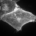
Hela cells transiently transfected with EGFP-GL-GPI show the organization of the membrane thru the distribution of GPI anchored protein. This image is the maximum z projection image that accompanies...
CIL:25359
NCBI Organism Classification
Canis lupus familiaris
Biological Process
cell extrusion
Cellular Component
cell
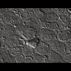
Reorganization of cells around a site of cell extrusion monitored live by time-lapse differential interference contrast microscopy. The process of extrusion began at hour 1 of observation and continu...
CIL:12410
NCBI Organism Classification
Cricetulus griseus
Biological Process
intracellular motility
Cellular Component
myosin IIA
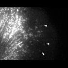
Localization and intracellular motility of myosin IIA (MIIA) in protrusive regions of migrating CHO.K1 cells. Arrowheads point to locations where representative MIIA filaments form. This Video corres...
CIL:12412
NCBI Organism Classification
Cricetulus griseus
Biological Process
intracellular motility
Cellular Component
myosin IIA/B
Localization of MIIA/B (head, actin-binding domain of MIIA (myosin IIA), and tail domain of MIIB (myosin IIB)) in MIIA-deficient CHO.K1 cells. This Video corresponds to Fig. 10 A and video 5 from JC...
CIL:12413
NCBI Organism Classification
Cricetulus griseus
Biological Process
intracellular motility
Cellular Component
myosin IIA/B
Localization of MIIB/A (head, actin-binding domain of MIIA (myosin IIA), and tail domain of MIIB (myosin IIB)) in MIIA-deficient CHO.K1 cells. This Video corresponds to Fig. 10 B and video 6 from CB 1...
CIL:11841
NCBI Organism Classification
Potorous tridactylus
Biological Process
microtubule polymerization
Cellular Component
microtubule
Microtubules in a Rac1(Q61L)-expressing PtK1 cell injected with Pak inhibitory fragment PBD/ID(H83L). Microtubules do not exhibit retrograde flow and undergo very little net growth. Epifluorescence i...
CIL:11845
NCBI Organism Classification
Potorous tridactylus
Biological Process
actin polymerization or depolymerization
Cellular Component
actin cytoskeleton
Actin dynamics in a Rac1(Q61L)-expressing PtK1 cell injected with Pak inhibitory fragment PBD/ID(H83L). Fast retrograde flow still occurs in the lamellipodium, but the width of the lamellipodium is re...
CIL:42163
NCBI Organism Classification
Homo sapiens
Biological Process
dissemination
Cellular Component
cell surface
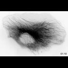
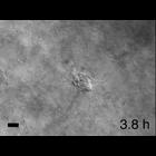
Time-lapse movie of a human tumor fragment grown in Matrigel that was freed and reembedded in collagen I. Images collected every 15 minutes and bar is 50 microns. CIL 42162 - CIL 42165 are related m...
CIL:42165
NCBI Organism Classification
Homo sapiens
Biological Process
dissemination
Cellular Component
cell surface
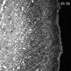
Time-lapse movie of a human tumor fragment grown in colllagen I that was freed and reembedded in collagen I. Images collected every 15 minutes and bar is 50 microns. CIL 42162 - CIL 42165 are relate...
CIL:40407
NCBI Organism Classification
none specified
Biological Process
cytoskeleton organization
Cellular Component
mitochondrion
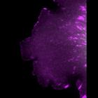
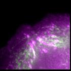
A confocal image of dividing keratinocyte cells labelled with mitotracker (red), tubulin (green) and nuclei (Dapi-blue). Cells were part of a study of the effect of Papilloma viruses in keratinocytes.
« Previous
1
...
20
21
22
23
24
25
26
27
...
237
Next »
Results per page:
10
20
50
100