Alternate header for print version
Advanced search
Contributors
Help
Submit
Search
menu
Cell Process
Cell Component
Cell Type
Organism
Microbial
Alzheimer's
Data Sets
Center for Research in Biological Systems
University of California, San Diego
9500 Gilman Drive
La Jolla, CA 92093-0608, USA
Voice
: (858) 534-0276
Fax
: (858) 534-7497
Email
: dorloff@ncmir.ucsd.edu
Search Results for
scanning electron microscopy
(448 results)
(Not the results you were expecting?)
(Comments)
Still Images
Video/Animation
Z-Stack
Time Series
CIL:40343
NCBI Organism Classification
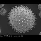
sildalcea malviflora
Biological Process
pollen wall assembly
Cellular Component
pollen wall
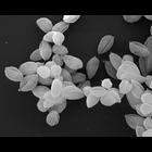
Scanning electron micrograph of sildalcea malviflora (prairie hollyhock) pollen. These specimens have been acetolyzed to remove cytoplasm and pollenkit in order to reveal the intricate wall structur...
CIL:40350
NCBI Organism Classification
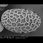
Lilium auratum
Biological Process
pollen wall assembly
Cellular Component
pollen wall
Scanning electron micrograph of Lilium auratum (Oriental Lily) pollen. These specimens have been acetolyzed to remove cytoplasm and pollenkit in order to reveal the intricate wall structure. This is p...
CIL:40355
NCBI Organism Classification
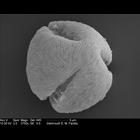
Rosa rugosa
Biological Process
pollen wall assembly
Cellular Component
pollen wall
Scanning electron micrograph of Rosa rugosa (Beach rose) pollen. These specimens have been acetolyzed to remove cytoplasm and pollenkit in order to reveal the intricate wall structure. This is part of...
CIL:42801
NCBI Organism Classification
none specified
Biological Process
none specified
Cellular Component
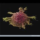
cell surface
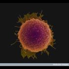
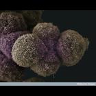
Colorized scanning electron micrograph of a cell cultured lung cancer cell. This image is part of an image group, CIL 42801-42803, showing several colorized scanning electron micrographs of cell cult...
CIL:42802
NCBI Organism Classification
none specified
Biological Process
none specified
Cellular Component
cell surface
Colorized scanning electron micrograph of a cell cultured lung cancer cell. This image is part of an image group, CIL 42801-42803, showing several colorized scanning electron micrographs of cell cult...
CIL:42803
NCBI Organism Classification
none specified
Biological Process
none specified
Cellular Component
cell surface
Colorized scanning electron micrograph of a cell cultured lung cancer cell. This image is part of an image group, CIL 42801-42803, showing several colorized scanning electron micrographs of cell cult...
CIL:12403
NCBI Organism Classification
Leishmania mexicana
Biological Process
amastigote form Leishmania mexicana
Cellular Component
none specified
Leishmania mexicana amastigote forms (WHO strain MNYC/BZ/62/M379). Amastigotes were grown in axenic culture. The cells were fixed with glutaraldehyde, dehydrated in ethanol, critical-point dried and s...
CIL:38811
NCBI Organism Classification
none specified
Biological Process
none specified
Cellular Component
cell surface
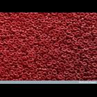
Scanning electron micrograph of red blood cells clearly showing their biconcave disc shape. Human red blood cells are typically 8 microns x 2 microns in size.
CIL:39104
NCBI Organism Classification
none specified
Biological Process
cell-cell adhesion
Cellular Component
cell surface
Scanning electron micrograph of pancreatic cancer cells. See additional image at CIL:39076.
CIL:39106
NCBI Organism Classification
none specified
Biological Process
none specified
Cellular Component
cell surface
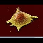
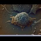
Colorized scanning electron micrograph of a lung cancer cell.
« Previous
1
...
8
9
10
11
12
13
14
15
...
45
Next »
Results per page:
10
20
50
100