Alternate header for print version
Advanced search
Contributors
Help
Submit
Search
menu
Cell Process
Cell Component
Cell Type
Organism
Microbial
Alzheimer's
Data Sets
Center for Research in Biological Systems
University of California, San Diego
9500 Gilman Drive
La Jolla, CA 92093-0608, USA
Voice
: (858) 534-0276
Fax
: (858) 534-7497
Email
: dorloff@ncmir.ucsd.edu
Search Results for
cone
(123 results)
(Not the results you were expecting?)
(Comments)
Still Images
Video/Animation
Z-Stack
Time Series
CIL:10115
NCBI Organism Classification
Rattus
Biological Process
developmental process
Cellular Component
cytoskeleton
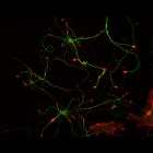
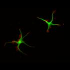
This multi-layer image shows the spatial relationship between filamentous actin (red) and microtubule array (green) in cultured hippocampal neurons, grown for 5 days in vitro. Actin staining (with rh...
CIL:10117
NCBI Organism Classification
Rattus
Biological Process
developmental process
Cellular Component
cytoskeleton
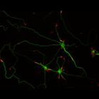
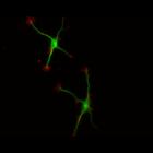
This multi-layer image shows the spatial relationship between filamentous actin (red) and microtubule array (green) in cultured hippocampal neurons, grown for 5 days in vitro. Actin staining (with rh...
CIL:10118
NCBI Organism Classification
Rattus
Biological Process
developmental process
Cellular Component
cytoskeleton
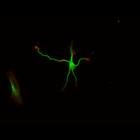
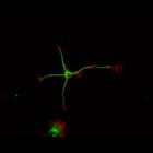
This multi-layer image shows the spatial relationship between filamentous actin (red) and microtubule array (green) in cultured hippocampal neurons, grown for 5 days in vitro. Actin staining (with rh...
CIL:10204
NCBI Organism Classification
Rattus
Biological Process
developmental process
Cellular Component
cytoskeleton
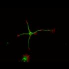
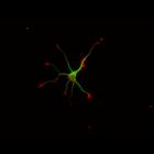
This multi-layer image shows the spatial relationship between filamentous actin (red) and microtubule array (green) in cultured hippocampal neurons, grown for 1 day in vitro. Actin staining (with rho...
CIL:10205
NCBI Organism Classification
Rattus
Biological Process
developmental process
Cellular Component
cytoskeleton
This multi-layer image shows the spatial relationship between filamentous actin (red) and microtubule array (green) in cultured hippocampal neurons, grown for 1 day in vitro. Actin staining (with rho...
CIL:10207
NCBI Organism Classification
Rattus
Biological Process
developmental process
Cellular Component
cytoskeleton
This multi-layer image shows the spatial relationship between filamentous actin (red) and microtubule array (green) in cultured hippocampal neurons, grown for 1 day in vitro. Actin staining (with rho...
CIL:10214
NCBI Organism Classification
Rattus
Biological Process
developmental process
Cellular Component
cytoskeleton
This multi-layer image shows the spatial relationship between filamentous actin (red) and microtubule array (green) in cultured hippocampal neurons, grown for 1 day in vitro. Actin staining (with rho...
CIL:8777
NCBI Organism Classification
Rattus
Biological Process
developmental process
Cellular Component
cytoskeleton
This color combined image shows the spatial relationship between filamentous actin (red) and microtubule array (green) in cultured hippocampal neurons, grown for 1 day in vitro. Actin staining (with ...
CIL:8778
NCBI Organism Classification
Rattus
Biological Process
developmental process
Cellular Component
cytoskeleton
This color combined image shows the spatial relationship between filamentous actin (red) and microtubule array (green) in cultured hippocampal neurons, grown for 1 day in vitro. Actin staining (with ...
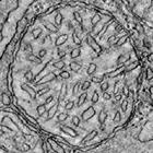
CIL:40169
NCBI Organism Classification
Mus musculus
Biological Process
none specified
Cellular Component
mitochondrion
Single tilt image (zero degree tilt) of a mitochondrion from a retinal rod photoreceptor of a female mouse (C57BL/6) from a 0.3 um section. This image has been downsampled from the raw data image whic...
« Previous
1
...
5
6
7
8
9
10
11
12
13
Next »
Results per page:
10
20
50
100