Alternate header for print version
Advanced search
Contributors
Help
Submit
Search
menu
Cell Process
Cell Component
Cell Type
Organism
Microbial
Alzheimer's
Data Sets
Center for Research in Biological Systems
University of California, San Diego
9500 Gilman Drive
La Jolla, CA 92093-0608, USA
Voice
: (858) 534-0276
Fax
: (858) 534-7497
Email
: dorloff@ncmir.ucsd.edu
Search Results for
Skeletal muscle contraction
(252 results)
(Not the results you were expecting?)
(Comments)
Still Images
Video/Animation
Z-Stack
Time Series
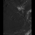
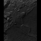
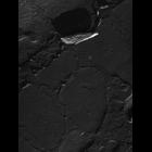
CIL:18488
NCBI Organism Classification
damselfly
Biological Process
skeletal muscle contraction
Cellular Component
myofibril
Most images show fractures through the outer membrane of mitochondria and SR/T tubules of dyads nestled in long grooves of the mitochondrial surface. All leaflets of the membranes appear in the image...
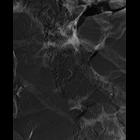
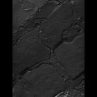
CIL:18489
NCBI Organism Classification
damselfly
Biological Process
skeletal muscle contraction
Cellular Component
myofibril
Most images show fractures through the outer membrane of mitochondria and SR/T tubules of dyads nestled in long grooves of the mitochondrial surface. All leaflets of the membranes appear in the image...
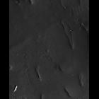
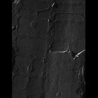
CIL:18491
NCBI Organism Classification
damselfly
Biological Process
skeletal muscle contraction
Cellular Component
myofibril
Most images show fractures through the outer membrane of mitochondria and SR/T tubules of dyads nestled in long grooves of the mitochondrial surface. All leaflets of the membranes appear in the image...
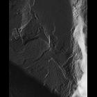
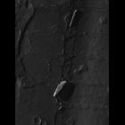
CIL:18493
NCBI Organism Classification
damselfly
Biological Process
skeletal muscle contraction
Cellular Component
myofibril
Most images show fractures through the outer membrane of mitochondria and SR/T tubules of dyads nestled in long grooves of the mitochondrial surface. All leaflets of the membranes appear in the image...
CIL:18455
NCBI Organism Classification
damselfly
Biological Process
skeletal muscle contraction
Cellular Component
myofibril
Most images show fractures through the outer membrane of mitochondria and SR/T tubules of dyads nestled in long grooves of the mitochondrial surface. All leaflets of the membranes appear in the image...
CIL:18460
NCBI Organism Classification
damselfly
Biological Process
skeletal muscle contraction
Cellular Component
myofibril
Most images show fractures through the outer membrane of mitochondria and SR/T tubules of dyads nestled in long grooves of the mitochondrial surface. All leaflets of the membranes appear in the image...
CIL:18462
NCBI Organism Classification
damselfly
Biological Process
skeletal muscle contraction
Cellular Component
myofibril
Most images show fractures through the outer membrane of mitochondria and SR/T tubules of dyads nestled in long grooves of the mitochondrial surface. All leaflets of the membranes appear in the image...
CIL:18463
NCBI Organism Classification
damselfly
Biological Process
skeletal muscle contraction
Cellular Component
myofibril
Most images show fractures through the outer membrane of mitochondria and SR/T tubules of dyads nestled in long grooves of the mitochondrial surface. All leaflets of the membranes appear in the image...
CIL:18464
NCBI Organism Classification
damselfly
Biological Process
skeletal muscle contraction
Cellular Component
myofibril
Most images show fractures through the outer membrane of mitochondria and SR/T tubules of dyads nestled in long grooves of the mitochondrial surface. All leaflets of the membranes appear in the image...
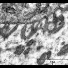
CIL:37224
NCBI Organism Classification
none specified
Biological Process
skeletal muscle contraction
Cellular Component
neuromuscular junction
Transmission electron micrograph of nerve cells at the thigh muscle motor end plate. The motor end plate is the highly-excitable region of muscle fiber plasma membrane responsible for initiation of a...
« Previous
1
...
12
13
14
15
16
17
18
19
...
26
Next »
Results per page:
10
20
50
100