Alternate header for print version
Advanced search
Contributors
Help
Submit
Search
menu
Cell Process
Cell Component
Cell Type
Organism
Microbial
Alzheimer's
Data Sets
Center for Research in Biological Systems
University of California, San Diego
9500 Gilman Drive
La Jolla, CA 92093-0608, USA
Voice
: (858) 534-0276
Fax
: (858) 534-7497
Email
: dorloff@ncmir.ucsd.edu
Search Results for
Louisa Howard
(136 results)
(Not the results you were expecting?)
(Comments)
Still Images
Video/Animation
Z-Stack
Time Series
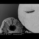
CIL:39384
NCBI Organism Classification
Amorphophallus titanum
Biological Process
pollen release
Cellular Component
anther
Transmission electron micrograph showing Amorphophallus titanum anther. The anther on the left is a cross section, showing the locule (cavity where the pollen is located). The anther on the right sh...
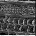
CIL:40380
NCBI Organism Classification
Solenostemon scutellarioides
Biological Process
plant stem organization
Cellular Component
cortex
Scanning electron microscope image of cross-section through a Solenostemon scutellarioidesi (coleus) stem. The image shows the outer epidermal layer, cortex, vascular bundles in a ring, and a central ...
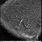
CIL:40385
NCBI Organism Classification
Oryza sativa
Biological Process
plant stem organization
Cellular Component
starch grain
Scanning electron microscope image of longitudinal section of Oryza sativa (rice) stem. Image shows cuts through the vascular cells and cortex cells. Starch granules are visible inside the vascular ce...
CIL:40388
NCBI Organism Classification
Psidium guajava
Biological Process
epidermis morphogenesis
Cellular Component
trichome
Scanning electron microscope image of Psidium guajava epidermal surface of the stem, showing trichomes (hair-like projections). This image is part of a group on botanical stems (CIL:40378-40395).
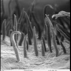
CIL:40392
NCBI Organism Classification
Helianthus annuus
Biological Process
epidermis morphogenesis
Cellular Component
trichome
Scanning electron microscope image of Helianthus annuus (sunflower) stem epidermal surface, showing a trichome (finger-like projection) and a stomata (pore used for gas exchange). This image is part ...
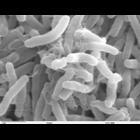
CIL:40397
NCBI Organism Classification
Vibrio cholerae
Biological Process
bacterial cell surface binding
Cellular Component
cell surface
Scanning electron microscope image of Vibrio cholerae bacteria, which infect the digestive system. This image is part of an image group that includes CIL:40395-40399.
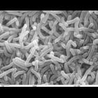
CIL:40398
NCBI Organism Classification
Vibrio cholerae
Biological Process
bacterial cell surface binding
Cellular Component
cell surface
Scanning electron microscope image of Vibrio cholerae bacteria, which infect the digestive system. This image is part of an image group that includes CIL:40395-40399.
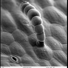
CIL:41307
NCBI Organism Classification
Penta lanceolata
Biological Process
pollen adhesion
Cellular Component
pollen coat
Scanning electron micrograph showing Penta lanceolata unacetolyzed pollen. Compare this pollen to acetolyzed pollen images by the same contributor in the Library. This image is a ]higher magnificatio...
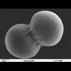
CIL:39782
NCBI Organism Classification
Lytechinus pictus
Biological Process
embryonic cleavage
Cellular Component
cleavage furrow
Scanning electron microscope image of Lytechinus pictus [sea urchin] embryo at the 2-cell stage. The fertilization envelope has been removed to reveal the cells covered with a dense meshwork of the hy...
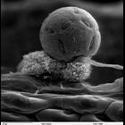
CIL:39790
NCBI Organism Classification
Lytechinus pictus
Biological Process
embryonic morphogenesis
Cellular Component
cell surface
Scanning electron microscope image of Strongylocentrotus drobachiensus [sea urchin] gastrula. This is a higher magnification image of CIL39765. The embryo was split open to reveal a nice cross-section...
« Previous
1
...
6
7
8
9
10
11
12
13
14
Next »
Results per page:
10
20
50
100