Alternate header for print version
Advanced search
Contributors
Help
Submit
Search
menu
Cell Process
Cell Component
Cell Type
Organism
Microbial
Alzheimer's
Data Sets
Center for Research in Biological Systems
University of California, San Diego
9500 Gilman Drive
La Jolla, CA 92093-0608, USA
Voice
: (858) 534-0276
Fax
: (858) 534-7497
Email
: dorloff@ncmir.ucsd.edu
Search Results for
Gregory Antipa
(65 results)
(Not the results you were expecting?)
(Comments)
Still Images
Video/Animation
Z-Stack
Time Series
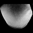
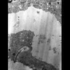
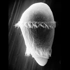
CIL:22783
NCBI Organism Classification
uncultured scuticociliate
Biological Process
cortical cytoskeleton organization
Cellular Component
cell cortex
A view of the left side of Conchophthirus curtus deciliated by a calcium shock method to reveal the organization of the ciliature of this densely ciliated organism. In particular this method reveals t...
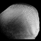
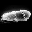
CIL:40905
NCBI Organism Classification
uncultured scuticociliate
Biological Process
cortical cytoskeleton organization
Cellular Component
cilium
Conchophthirus was deciliated by calcium ion shock followed by shearing through a micropipette. This revealed the location of the deciliated basal bodies and the cell's surface architecture. In this i...
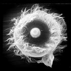
CIL:39251
NCBI Organism Classification
Didinium nasutum
Biological Process
ciliary cell motility
Cellular Component
oral apparatus
Didinium nasutum showing the two prominent girdles of cilia in full metachronous beat. If you look at higher magnification you will see the occasional short, blunt clavate cilia. These are known to no...
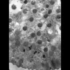
CIL:15470
NCBI Organism Classification
Didinium nasutum
Biological Process
macronucleus organization
Cellular Component
macronucleus
Food vacuole and macronucleus of Didinium. The food vacuole contains unexploded trichocysts of Paramecium. Further details available at Wessenberg, H. and Antipa, G. 1968. Studies on Didinium nasutum....
CIL:15713
NCBI Organism Classification
Didinium nasutum
Biological Process
contractile vacuole organization
Cellular Component
contractile vacuole
The contractile vacuole and cortex of Didinium nasutum showing an extrusive organelle (cyrtocyst) of unknown function. Further details available at Wessenberg, H. and Antipa, G. 1968. Studies on Didin...
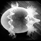
CIL:19536
NCBI Organism Classification
Didinium nasutum
Biological Process
oral apparatus organization
Cellular Component
oral apparatus
Apical oral view of Didinium. Showing the proboscis containing extrusive organelles to demobilize and eventually engulph it's prey. Metachronous waves of cilia in the anterior girdle of the two charac...
CIL:15400
NCBI Organism Classification
Didinium nasutum
Biological Process
oral apparatus organization
Cellular Component
oral apparatus
A cross section of the probosis oral apparatus of Didinium showing the organization, distribution of toxicysts, and the ribbons of microtubules that support the ingestion of large prey. Further detail...
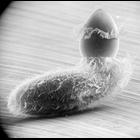
CIL:22782
NCBI Organism Classification
Didinium nasutum
Biological Process
phagocytosis
Cellular Component
oral apparatus
Didinium intiates capture of Paramecium. Believe-it-or-not, this Didinium may accomplish the ingestion of the Paramecium in less than one minute. See other images in the link below. Also showing metac...
CIL:21992
NCBI Organism Classification
Didinium nasutum
Biological Process
phagocytosis
Cellular Component
oral apparatus
Didinium ingests Paramecium. Note that Paramecium is nearly the waiting food vacuole and the proboscis of Didinium is beginning to reform. This micrograph also shows the metachronous waves of cilia in...
CIL:21995
NCBI Organism Classification
Didinium nasutum
Biological Process
phagocytosis
Cellular Component
oral apparatus
Didinium ingests Paramecium. Note that Paramecium is folded in half as it is compressed and enters the waiting food vacuole. This micrograph also shows a few discharged Paramecium trichocysts as well ...
« Previous
1
2
3
4
5
6
7
Next »
Results per page:
10
20
50
100