Alternate header for print version
Advanced search
Contributors
Help
Submit
Search
menu
Cell Process
Cell Component
Cell Type
Organism
Microbial
Alzheimer's
Data Sets
Center for Research in Biological Systems
University of California, San Diego
9500 Gilman Drive
La Jolla, CA 92093-0608, USA
Voice
: (858) 534-0276
Fax
: (858) 534-7497
Email
: dorloff@ncmir.ucsd.edu
Search Results for
Dieter Brandner; Ginger Withers
(283 results)
(Not the results you were expecting?)
(Comments)
Still Images
Video/Animation
Z-Stack
Time Series
CIL:6138
NCBI Organism Classification
Rattus
Biological Process
neuron development
Cellular Component
cytoskeleton
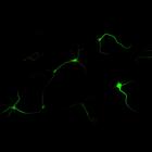
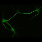
Synapse formation in cultured hippocampal neurons after 7 days in vitro. Cells were immunostained for MAP2, a microtubule associated protein localized to dendrites but not axons (green), and synapsin...
CIL:10095
NCBI Organism Classification
Rattus
Biological Process
developmental process
Cellular Component
cytoskeleton
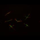
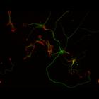
This multi-layer image shows the spatial relationship between filamentous actin (red) and microtubule array (green) in cultured hippocampal neurons, grown for 1 day in vitro. Actin staining (with rho...
CIL:10096
NCBI Organism Classification
Rattus
Biological Process
developmental process
Cellular Component
cytoskeleton
This multi-layer image shows the spatial relationship between filamentous actin (red) and microtubule array (green) in cultured hippocampal neurons, grown for 1 day in vitro. Actin staining (with rho...
CIL:10109
NCBI Organism Classification
Rattus
Biological Process
developmental process
Cellular Component
cytoskeleton
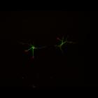
This multi-layer image shows the spatial relationship between filamentous actin (red) and microtubule array (green) in cultured hippocampal neurons, grown for 3 days in vitro. Actin staining (with rh...
CIL:10112
NCBI Organism Classification
Rattus
Biological Process
developmental process
Cellular Component
cytoskeleton
This multi-layer image shows the spatial relationship between filamentous actin (red) and microtubule array (green) in cultured hippocampal neurons, grown for 3 days in vitro. Actin staining (with rh...
CIL:10264
NCBI Organism Classification
Rattus
Biological Process
cellular developmental process
Cellular Component
dendrite
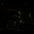
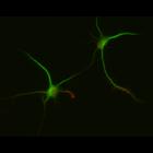
Early stages of dendritic development and synapse formation in cultured hippocampal neurons. This multilayer image shows neurons fixed at 7 days in vitro and immunostained for the dendritically loca...
CIL:10267
NCBI Organism Classification
Rattus
Biological Process
cellular developmental process
Cellular Component
dendrite
Early stages of dendritic development and synapse formation in cultured hippocampal neurons. This multilayer image shows neurons fixed at 7 days in vitro and immunostained for the dendritically loca...
CIL:10268
NCBI Organism Classification
Rattus
Biological Process
cellular developmental process
Cellular Component
dendrite
Early stages of dendritic development and synapse formation in cultured hippocampal neurons. This multilayer image shows neurons fixed at 7 days in vitro and immunostained for the dendritically loca...
CIL:10312
NCBI Organism Classification
Rattus
Biological Process
cellular developmental process
Cellular Component
dendrite
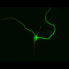
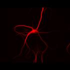
Cultured hippocampal neurons after 10 days in vitro, immunostained for MAP2, a microtubule associated protein localized to dendrites (red) but not axons, which are not apparent in the immunofluorescen...
CIL:10315
NCBI Organism Classification
Rattus
Biological Process
cellular developmental process
Cellular Component
dendrite
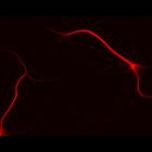
Cultured hippocampal neurons after 10 days in vitro, immunostained for MAP2, a microtubule associated protein localized to dendrites (red) but not axons, which are not apparent in the immunofluorescen...
« Previous
1
...
9
10
11
12
13
14
15
16
...
29
Next »
Results per page:
10
20
50
100