Alternate header for print version
Advanced search
Contributors
Help
Submit
Search
menu
Cell Process
Cell Component
Cell Type
Organism
Microbial
Alzheimer's
Data Sets
Center for Research in Biological Systems
University of California, San Diego
9500 Gilman Drive
La Jolla, CA 92093-0608, USA
Voice
: (858) 534-0276
Fax
: (858) 534-7497
Email
: dorloff@ncmir.ucsd.edu
Search Results for
147
(31 results)
(Not the results you were expecting?)
(Comments)
Still Images
Video/Animation
Z-Stack
Time Series
CIL:147
NCBI Organism Classification
Potorous tridactylus
Biological Process
none specified
Cellular Component
cell
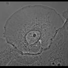
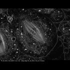
Phase contrast image of a PTK-1 cell expressing constitutively active Rac1(Q61L). Live cells were visualized using X-rhodamine tubulin (injected at 1 mg/mL)and eGFP-Rac1(Q61L) (injected into the nucl...
CIL:11050
NCBI Organism Classification
Cavia porcellus
Biological Process
nucleus organization
Cellular Component
nuclear envelope
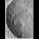
Transmission electron micrograph of surface of nuclear envelope of guinea pig spermatocyte prepared by the Freeze-fracture technique shows the unusual clustering of nuclear pores in these cells. From...
CIL:11048
NCBI Organism Classification
Cavia porcellus
Biological Process
nucleus organization
Cellular Component
nuclear envelope
Nuclear pores are clearly seen in these transmission electron micrographs of cells after cryo-fixation, freeze-fracture and surface replication. Upper panel shows the outer surface of of the nuclear e...
CIL:8060
NCBI Organism Classification
Rattus norvegicus
Biological Process
secretion
Cellular Component
Golgi apparatus part
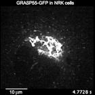
Traffic Jam: The Dynamic Behavior of a Golgi Matrix Protein This video shows an NRK cell stably expressing the Golgi matrix protein GRASP55-GFP. The Golgi apparatus in mammalian cells is made up of ...
CIL:35284
NCBI Organism Classification
Lilium formosanum
Biological Process
pollen tube growth
Cellular Component
mitochondria
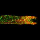
Living Lilium pollen tube labeled with mitotracker green to mark mitochondria (green) and cytochalasin D TRITC to mark endoplasmic reticulum (red) imaged using laser scanning focal microscopy. Shown i...
CIL:11113
NCBI Organism Classification
Oncorhynchus kisutch
Biological Process
plasma membrane organization
Cellular Component
cell surface
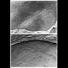
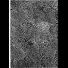
Scanning electron micrograph of the epidermal surface of the Pacific coho salmon. Ridge-like folds of the plasmalemma, called microplicae, display a labyrinth pattern across the surface, and a circum...
CIL:12025
NCBI Organism Classification
Nephrotoma
Biological Process
male meiosis chromosome segregation
Cellular Component
spindle
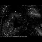
Movie of meiosis I in primary spermatocytes of the crane-fly Nephrotoma suturalis that have been experimentally flattened. Time-lapse polarization microscopy using a Nikon Microphot SA, equipped for...
CIL:12373
NCBI Organism Classification
Nephrotoma
Biological Process
male meiosis chromosome segregation
Cellular Component
spindle
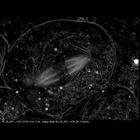
Movie showing the dynamics of kinetchore microtubules during meiosis I in primary spermatocytes of the crane-fly Nephrotoma suturalis that were experimentally flattened. Time-lapse polarization micros...
CIL:12028
NCBI Organism Classification
Nephrotoma
Biological Process
male meiosis chromosome segregation
Cellular Component
spindle
Time series of meiosis I, starting at metaphase, in primary spermatocytes of the crane-fly Nephrotoma suturalis that have been experimentally flattened. Time-lapse polarization microscopy using a Nik...
CIL:11046
NCBI Organism Classification
Cavia sp.
Biological Process
nucleus organization
Cellular Component
nuclear pore
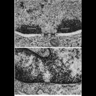
Transmission electron micrographs showing details of nuclear pores in thin sections of an erythroblast (above) and endothelial cell (below). Pores appear to be filled with dark staining material in th...
1
2
3
4
Next »
Results per page:
10
20
50
100