Alternate header for print version


Licensing
Public Domain: This image is in the public domain and thus free of any copyright restrictions. However, as is the norm in scientific publishing and as a matter of courtesy, any user should credit the content provider for any public or private use of this image whenever possible. Learn more
tweet  
*CIL – Cell Image Library accession number. Please use this to reference an image.

CIL:12028*  Cite 
Description

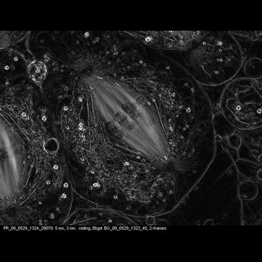
Time series of meiosis I, starting at metaphase, in primary spermatocytes of the crane-fly Nephrotoma suturalis that have been experimentally flattened. Time-lapse polarization microscopy using a Nikon Microphot SA, equipped for liquid crystal polarized light microscopy (LC-PolScope, CRi, Woburn Massachusetts) 60x/1.4 PlanApo oil immersion objective, 1.4 NA oil immersion condenser, with 2.0x zoom lens. Images captured every 15 sec over 35 min using a QImaging Retigo EXi CCD camera. Raw images were processed using 5-frame algorithm (Shribak and Oldenbourg, 2003). A movie of the series is included in this grouped set.

Biological Sources
NCBI Organism Classification
Nephrotoma
Nephrotoma suturalis
Cell Type
primary spermatocyte
Cellular Component
spindle
nuclear chromosome
Biological Context
Biological Process
male meiosis chromosome segregation
Attribution
Names
James R. LaFountain
Rudolf Oldenbourg
Published
LaFountain JR, et al. (2004) Mol Cell Biol 15, 5724-5732
Published
Oldenbourg R, Mei G (1995) J Microsc 180, 140-147.
Published
Shribak M, Oldenbourg R (2003) Appl Opt 42, 3009-3017
Pubmed
15469981
Pubmed
8537959
Pubmed
12790452
Citation
Digital Object Identifier (DOI)
doi:10.7295/W9CIL12028
Archival Resource Key (ARK)
ark:/b7295/w9cil12028
Grouping This image is part of a group.
Imaging
Image Type
charge coupled device (CCD)
Image Mode
polarization microscopy
Parameters Imaged
UV/visible/IR photons
Source of Contrast
intrinsic birefringence
Visualization Methods
relative retardation (retardance) measurement
Processing History
5-frame algorithm
Sample Preparation
Methods
living tissue
Relation To Intact Cell
whole mounted tissue
Dimensions
Spatial Axis Image Size Pixel Size
X 678px 87nm
Y 518px 87nm
Time 15 seconds 150