Alternate header for print version
Advanced search
Contributors
Help
Submit
Search
menu
Cell Process
Cell Component
Cell Type
Organism
Microbial
Alzheimer's
Data Sets
University of California, San Diego
9500 Gilman Drive
La Jolla, CA 92093-0608, USA
Voice
: (858) 534-0276
Fax
: (858) 534-7497
Email
: dorloff@ncmir.ucsd.edu
Search Results for
electron-dense stain
(2012 results)
(Not the results you were expecting?)
(Comments)
CIL:10440
NCBI Organism Classification
Rana catesbeiana
Biological Process
intermediate filament cytoskeleton organization
Cellular Component
intermediate filament cytoskeleton
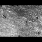
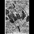
This group of micrographs illustrate the ultrastructural characteristics of the middle layer of the three meningeal layers that cover the central nervous system The arachnoid cells contain abundant in...
CIL:11054
NCBI Organism Classification
Anura
Biological Process
nucleus organization
Cellular Component
nuclear envelope
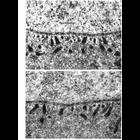
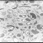
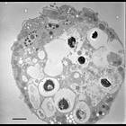
Transmission electron micrographs of sections of tadpole oocyte show the potential for exchange between the nucleus and the cytoplasm at the nuclear envelope. Thin filaments that appear to traverse nu...
CIL:11056
NCBI Organism Classification
Hirudinea
Biological Process
nucleus organization
Cellular Component
nuclear envelope
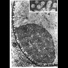
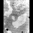
Transmission electron micrograph illustrating the unusually prominent fibrous nuclear lamina (arrows) seen in many invertebrates such as this leech ganglion cell. From Fawcett (1966) Am J. Anat. 119:...
CIL:11643
NCBI Organism Classification
Carassius sp. U8MN
Biological Process
stereocilium organization
Cellular Component
stereocilium
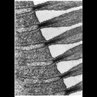
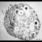
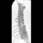
Figure 327 from Chapter 13 (Cilia and Flagella) of 'The Cell' by Don W. Fawcett M.D. Longitudinal section of the apex of a hair cell from the saccular macula of the goldfish. The stereocilia are seen ...
CIL:11556
NCBI Organism Classification
Gallus gallus
Biological Process
spermatogonial cell division
Cellular Component
pericentriolar material
Figure 302 from Chapter 12 (Centrioles) by Don Fawcett. The centrioles replicate early in cell division and take up positions at either pole of the division figure. Concurrently with the condensation ...
CIL:12311
NCBI Organism Classification
Nassula
Biological Process
cytoplasm organization
Cellular Component
cytoplasm
Detail of cytopharyngeal vesicles. TEM taken on 3/2/71 by R. Allen with Hitachi HU11A operating at 75kV. Neg. 19,500X. Bar = 0.5µm. A print of the negative was scanned and processed in Photoshop. Th...
CIL:12312
NCBI Organism Classification
Nassula
Biological Process
contractile vacuole organization
Cellular Component
contractile vacuole
Contractile vacuole of Nassula sp. Parts of the single CV appear on two sides of the CV pore. Fields of spongiome abut the filled portions of the CV. TEM taken on 3/27/69 by R. Allen with Philips 300 ...
CIL:12324
NCBI Organism Classification
Halteria grandinella
Biological Process
macronucleus organization
Cellular Component
macronucleus
This group represents a serial series of sections. At the level of this section the macronucleus, cytopharyngeal tube, food vacuoles, and perioral membranelle are evident. TEM taken on 3/12/71 by R. A...
CIL:12327
NCBI Organism Classification
Halteria grandinella
Biological Process
macronucleus organization
Cellular Component
macronucleus
This group represents a serial series of sections. At the level of this section, the bottom of cytopharyngeal tube with one cilium remaining. End of macronucleus, three membranelles of the pek, and ma...
CIL:12337
NCBI Organism Classification
Euplotes sp.
Biological Process
oral apparatus organization
Cellular Component
oral apparatus
The transition from 3 to 2 rows of cilia can be seen in this view of the AZM. TEM taken on 7/24/67 by R. Allen with Philips 200 operating at 60kV. Neg. 1,370X. Bar = 5µm. Standard glutaraldehyde fix...
« Previous
1
...
14
15
16
17
18
19
20
21
...
202
Next »
Results per page:
10
20
50
100