Alternate header for print version


Licensing
Attribution Non-Commercial; No Derivatives:This image is licensed under a Creative Commons Attribution, Non-Commercial, No Derivatives License. View License Deed | View Legal Code
tweet  
*CIL – Cell Image Library accession number. Please use this to reference an image.

CIL:11643*  Cite 
Description

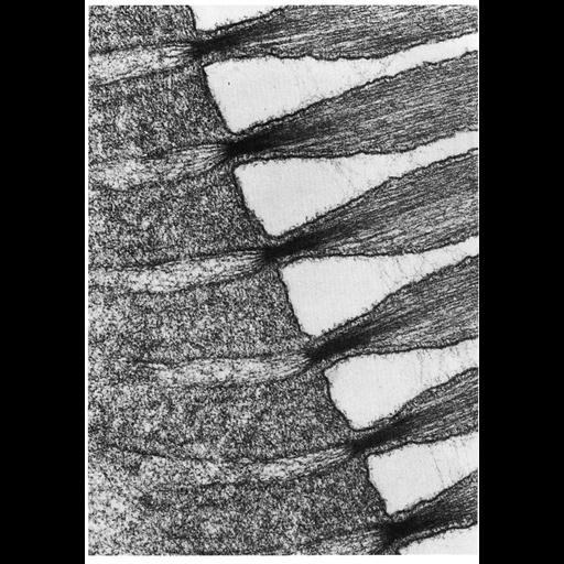
Figure 327 from Chapter 13 (Cilia and Flagella) of 'The Cell' by Don W. Fawcett M.D. Longitudinal section of the apex of a hair cell from the saccular macula of the goldfish. The stereocilia are seen in longitudinal section, the sensory hairs are narrower at their base and contain a large number of parallel filaments that converge into a dense bundle in the narrow basal region of the hair and then diverge again as they continue into the cuticular plate in the apical cytoplasm. A copy of the chapter is available on the ASCB's BioEDUCATE website.

Biological Sources
NCBI Organism Classification
Carassius sp. U8MN
Cell Type
saccular macula
Cellular Component
stereocilium
Biological Context
Biological Process
stereocilium organization
auditory receptor cell stereocilium organization
Attribution
Names
Don W. Fawcett
Kogaku Saito
Kiyoshi Hama
Published
ISBN 0721635849
Link
http://www.ascb.org/bioeducate/FawcettTheCell...
Citation
Digital Object Identifier (DOI)
doi:10.7295/W9CIL11643
Archival Resource Key (ARK)
ark:/b7295/w9cil11643
Grouping This image is part of a group.
Imaging
Image Type
transmission electron microscopy (TEM)
illumination by electrons
Image Mode
detection of electrons
film
Parameters Imaged
electron density
Source of Contrast
stain with broad specificity
Visualization Methods
stain with broad specificity
osmium tetroxide
uranyl salt
lead salt
Processing History
recorded image
film
Print from negative scanned for Photoshop.
Data Qualifiers
processed data
Dimensions
Spatial Axis Image Size Pixel Size
X 898px ——
Y 1283px ——