Alternate header for print version
Advanced search
Contributors
Help
Submit
Search
menu
Cell Process
Cell Component
Cell Type
Organism
Microbial
Alzheimer's
Data Sets
University of California, San Diego
9500 Gilman Drive
La Jolla, CA 92093-0608, USA
Voice
: (858) 534-0276
Fax
: (858) 534-7497
Email
: dorloff@ncmir.ucsd.edu
Search Results for
detection of electrons
(1490 results)
(Not the results you were expecting?)
(Comments)
CIL:9976
NCBI Organism Classification
notopthalmus viridescens
Biological Process
DNA packaging
Cellular Component
nuclear chromatin
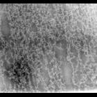
Newt (Notopthalmus viridescens erythrocytes were isolated in 0.1M KCl, spread on the surface of Na-citrate, picked up on formvar-carbon films, fixed with glutaraldehyde and paraformaldehyde and negati...
CIL:9978
NCBI Organism Classification
notopthalmus viridescens
Biological Process
DNA packaging
Cellular Component
nuclear chromatin
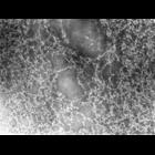
Newt (Notopthalmus viridescens erythrocytes were isolated in 0.1M KCl, spread on the surface of Na-citrate, picked up on formvar-carbon films, fixed with glutaraldehyde and paraformaldehyde and negati...
CIL:9984
NCBI Organism Classification
notopthalmus viridescens
Biological Process
DNA packaging
Cellular Component
nuclear chromatin
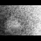
Newt (Notopthalmus viridescens erythrocytes were isolated in 0.1M KCl, spread on the surface of Na-citrate, picked up on formvar-carbon films, fixed with glutaraldehyde and paraformaldehyde and negati...
CIL:10944
NCBI Organism Classification
Homo sapiens
Biological Process
cell-substrate adhesion
Cellular Component
basal lamina
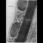
This electron micrograph shows the endothelium and the underlying Descemet's membrane of the human cornea. Descemet's membrane is an unusually thick and structurally specialized example of basal lami...
CIL:10987
NCBI Organism Classification
Unspecified
Biological Process
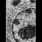
nucleus organization
Cellular Component
nucleus
Transmission electron micrograph of glutaraldehyde fixed pancreatic acinar cell showing the characteristic features of nuclear chromatin with this preparative method. Darkly staining heterochromatin (...
CIL:11014
NCBI Organism Classification
Ovis aries
Biological Process
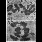
nucleus organization
Cellular Component
nuclear chromosome
Transmission electron micrographs showing chromosomes in dividing spermatogonia during metaphase in tissue from ram testis (upper panel) and as they begin to separate during early anaphase in tissue f...
CIL:11023
NCBI Organism Classification
Homo sapiens
Biological Process
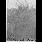
nucleus organization
Cellular Component
nuclear chromosome
Transmission electron micrograph showing part of a decondensed mitotic chromosome. The residual protein 'scaffold' (at bottom) is surrounded by a 'halo' of DNA loops 20 to 24 micrometers long. The en...
CIL:11225
NCBI Organism Classification
none specified
Biological Process
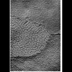
cell communication
Cellular Component
gap junction
Replica of a freeze-fractured gap junction presents the inner half-membrane on one of the cells (P-face, lower half of the micrograph) and the the outer half-membrane of the other cells (E-face, upper...
CIL:11364
NCBI Organism Classification
Rattus
Biological Process
post-translational protein modification
Cellular Component
Golgi apparatus
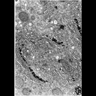
Figure200 from Chapter 6 (Golgi Apparatus) of 'The Cell, 2nd Ed.' by Don W. Fawcett M.D. The Golgi apparatus in epithelial cells of rat epididymis. The tissue was stained with thiamine pyrophosphata...
CIL:11370
NCBI Organism Classification
Cavia porcellus
Biological Process
post-translational protein modification
Cellular Component
Golgi apparatus
Figure 205 from Chapter 6 (Golgi Apparatus) of 'The Cell, 2nd Ed.' by Don W. Fawcett M.D. Sequence of the development of the acrosomal cap in spermatids from guinea pig testis, as observed in electro...
« Previous
1
...
29
30
31
32
33
34
35
36
...
149
Next »
Results per page:
10
20
50
100