Alternate header for print version


Licensing
Attribution Non-Commercial; No Derivatives:This image is licensed under a Creative Commons Attribution, Non-Commercial, No Derivatives License. View License Deed | View Legal Code
tweet  
*CIL – Cell Image Library accession number. Please use this to reference an image.

CIL:11225*  Cite 
Description

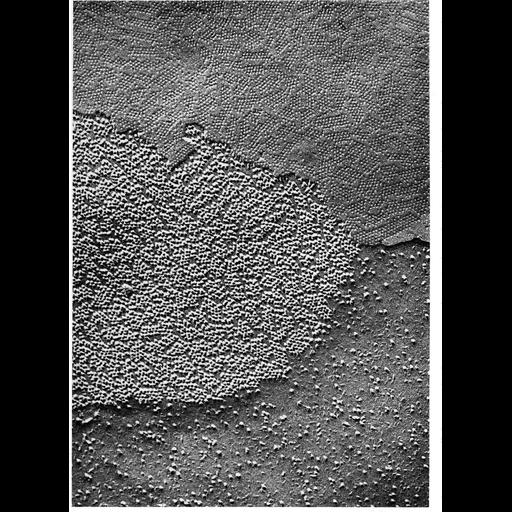
Replica of a freeze-fractured gap junction presents the inner half-membrane on one of the cells (P-face, lower half of the micrograph) and the the outer half-membrane of the other cells (E-face, upper part of the micrograph). Image by Daniel Friend is Figure 96 from Chapter 3 (Junctional Specializations) of 'The Cell, 2nd Ed.' by Don W. Fawcett M.D. A PDF copy of the accompanying chapter is available on the ASCB's BioEDUCATE website.

Biological Sources
Cellular Component
gap junction
connexon complex
plasma membrane
Biological Context
Biological Process
cell communication
cell-cell junction organization
Attribution
Names
Daniel Friend
Published
ISBN 0721635849
Link
The Cell, 2nd Ed. by D.W. Fawcett
Citation
Digital Object Identifier (DOI)
doi:10.7295/W9CIL11225
Archival Resource Key (ARK)
ark:/b7295/w9cil11225
Grouping This image is part of a group.
Imaging
Image Type
transmission electron microscopy (TEM)
Image Mode
illumination by electrons
detection of electrons
Parameters Imaged
electron density
Source of Contrast
differences in deposition of metal shadow
Visualization Methods
shadowing and plating
Data Qualifiers
processed data
Sample Preparation
Methods
freeze_fracture/freeze_etch
Relation To Intact Cell
isolated subcellular component
Dimensions
Spatial Axis Image Size Pixel Size
X 926px ——
Y 1268px ——