Alternate header for print version
Advanced search
Contributors
Help
Submit
Search
menu
Cell Process
Cell Component
Cell Type
Organism
Microbial
Alzheimer's
Data Sets
University of California, San Diego
9500 Gilman Drive
La Jolla, CA 92093-0608, USA
Voice
: (858) 534-0276
Fax
: (858) 534-7497
Email
: dorloff@ncmir.ucsd.edu
Search Results for
whole mounted tissue
(2808 results)
(Not the results you were expecting?)
(Comments)
CIL:220
NCBI Organism Classification
Mus musculus
Biological Process
none specified
Cellular Component
none specified
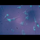
Molecules designed to block the expanded CAG repeats characteristic of Huntington's disease fluoresce blue in these cultured cells and prevent the expression of the mutant proteins that cause the di...
CIL:240
NCBI Organism Classification
none specified
Biological Process
none specified
Cellular Component
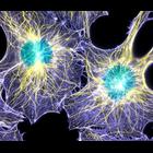
actin cytoskeleton
Two interphase cells with immunofluorescence labeling of actin filaments (purple), microtubules (yellow), and nuclei (green). This image won first place in the Nikon 2003 Small World photo competition...
CIL:27153
NCBI Organism Classification
unidentified plant
Biological Process
reproduction
Cellular Component
none specified
Z-stack through an unidentified pollen (mixed pollen grain w.m.; Carolina, commercial sample), with 200 image planes. See accompanying image in this group for a colorized maximum projection.
CIL:38954
NCBI Organism Classification
Ranunculus
Biological Process
water transport
Cellular Component
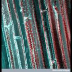
xylem vessels
A confocal micrograph of stained and autofluorescent cells of xylem in the stem of buttercup (Ranunculus), showing xylem vessels whose walls are studded with pits that allow passage of water from one ...
CIL:39086
NCBI Organism Classification
none specified
Biological Process
none specified
Cellular Component
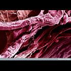
myofibril
Colorized scanning electron micrograph of thigh muscle fibrils.
CIL:39089
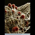
NCBI Organism Classification
Plasmodium yoelii nigeriensis
Biological Process
sporozoan zygote development
Cellular Component
cyst wall
Colorized scanning electron micrograph of malaria (Plasmodium yoelii nigeriensis) oocysts ( thick-walled structure in which sporozoan zygotes develop) developing on the midgut wall of the mosquito Ano...
CIL:39346
NCBI Organism Classification
Passiflora edulis
Biological Process
pollen wall organization
Cellular Component
pollen wall
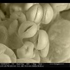
Scanning electron micrograph of passion flower pollen. A related image is CIL:39344.
CIL:41462
NCBI Organism Classification
diatom
Biological Process
none specified
Cellular Component
chlorophyll
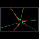
Star-like colonial diatom imaged with fluorescence microscopy. Green indicates newly depositing silica, red is chlorophyll. Tenth Prize, 2008 Olympus BioScapes Digital Imaging Competition®.
CIL:41569
NCBI Organism Classification
Elphidium crispum
Biological Process
cell surface organization
Cellular Component
cell surface
Brightfield image of protozoan Elphidium crispum found growing on the Dorset coast of England. Honorable Mention, 2011 Olympus BioScapes Digital Imaging Competition®.
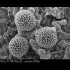
CIL:41616
NCBI Organism Classification
Galium odoratum
Biological Process
none specified
Cellular Component
pollen coat
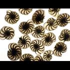
Colorized scanning electron micrograph of pollen grains that are believed to be from Galium Odoratum or "sweet woodruff", a small flower native to South Carolina.
« Previous
1
...
9
10
11
12
13
14
15
16
...
281
Next »
Results per page:
10
20
50
100
")