Alternate header for print version
Advanced search
Contributors
Help
Submit
Search
menu
Cell Process
Cell Component
Cell Type
Organism
Microbial
Alzheimer's
Data Sets
University of California, San Diego
9500 Gilman Drive
La Jolla, CA 92093-0608, USA
Voice
: (858) 534-0276
Fax
: (858) 534-7497
Email
: dorloff@ncmir.ucsd.edu
Search Results for
scanning electron microscopy (SEM)
(367 results)
(Not the results you were expecting?)
(Comments)
CIL:37315
NCBI Organism Classification
Cavia porcellus
Biological Process
none specified
Cellular Component
cell surface
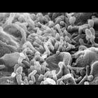
Scanning electron micrograph of single guinea pig pancreatic acinar cell. Image made available by James D. Jamieson and the Department of Cell Biology, Yale University School of Medicine.
CIL:38812
NCBI Organism Classification
none specified
Biological Process
ephrin B2 mutation
Cellular Component
none specified
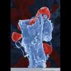
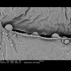
Color-enhanced scanning electron micrograph of red blood cells leaking out of a ruptured blood vessel. This is due to a mutation in the ephrin-B2 gene that causes the blood vessels to be more fragile ...
CIL:39088
NCBI Organism Classification
Mus musculus
Biological Process
liver trabeculae organization
Cellular Component
cell surface
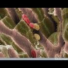
This scanning electron micrograph shows the internal structure of liver tissue from an adult mouse. The sinusoids (vascular channels lined with endothelial cells) can be seen as pink structures runnin...
CIL:38992
NCBI Organism Classification
Danio rerio
Biological Process
sensory perception
Cellular Component
stereocilium
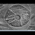
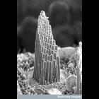
Scanning electron micrograph of the apical surface of neuromast organ of lateral line system in a 17-day-old zebrafish. The hair bundles at the top of each sensory cell emerge through a pore in the ...
CIL:39012
NCBI Organism Classification
Malaclemys terrapin
Biological Process
detection of mechanical stimulus involved in sensory perception of sound
Cellular Component
stereocilium bundle
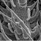
A scanning electron microscope image of the sensory hair bundle of a single hair cell from a terrapin's hearing organ in the inner ear. Vibrations made by sound cause the hairs to be moved back and fo...
CIL:39342
NCBI Organism Classification
Helianthus annuus
Biological Process
none specified
Cellular Component
none specified
Scanning electron microscope image of sunflower lower leaf surface where a vein is located. This is part of an image series containing images CIL:39341-39343, and 39345.
CIL:39381
NCBI Organism Classification
Amorphophallus titanum
Biological Process
pollen development
Cellular Component
pollen wall
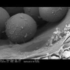
Transmission electron micrograph of an Amorphophallus titanum anther. This high magnification image shows pollen inside the locule (cavity where the pollen is located). This image is part of a group...
CIL:39382
NCBI Organism Classification
Amorphophallus titanum
Biological Process
pollen development
Cellular Component
pollen wall
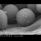
Transmission electron micrograph of an Amorphophallus titanum anther. This high magniification image shows pollen inside the locule (cavity where the pollen is located). This image is part of a grou...
CIL:39383
NCBI Organism Classification
Amorphophallus titanum
Biological Process
pollen release
Cellular Component
pollen wall
Transmission electron micrograph of an Amorphophallus titanum anther. This high magniification image shows pollen just being released from the opening at the top of the anther. This image is part of ...
CIL:39384
NCBI Organism Classification
Amorphophallus titanum
Biological Process
pollen release
Cellular Component
anther
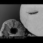
Transmission electron micrograph showing Amorphophallus titanum anther. The anther on the left is a cross section, showing the locule (cavity where the pollen is located). The anther on the right sh...
« Previous
1
...
7
8
9
10
11
12
13
14
...
37
Next »
Results per page:
10
20
50
100