Alternate header for print version


Licensing
Attribution Non-Commercial Share Alike:This image is licensed under a Creative Commons Attribution, Non-Commercial Share Alike License. View License Deed | View Legal Code
tweet  
*CIL – Cell Image Library accession number. Please use this to reference an image.

CIL:37315*  Cite 
Description

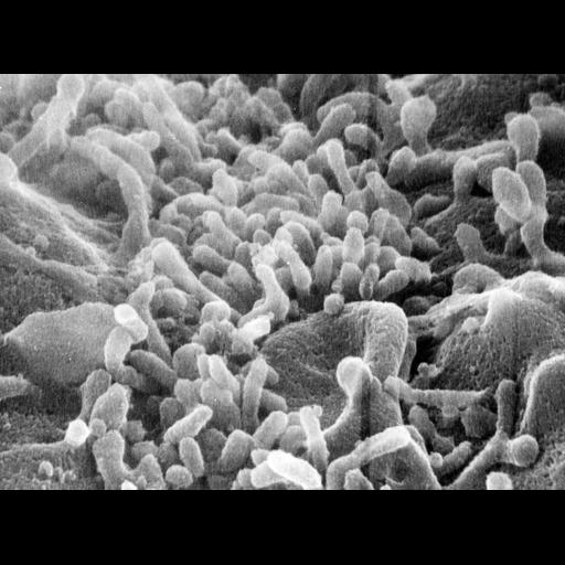
Scanning electron micrograph of single guinea pig pancreatic acinar cell. Image made available by James D. Jamieson and the Department of Cell Biology, Yale University School of Medicine.

Technical Details

Original 3.25 in. x 4 in. lantern slides were scanned at 600dpi. Original magnification x2,000.

Biological Sources
NCBI Organism Classification
Cavia porcellus
Cell Type
pancreatic acinar cell
Cellular Component
cell surface
Attribution
Names
James D Jamieson
A. Amsterdam
Link
George E. Palade EM Slide Colleection
Citation
Digital Object Identifier (DOI)
doi:10.7295/W9CIL37315
Archival Resource Key (ARK)
ark:/b7295/w9cil37315
Imaging
Image Type
recorded image
Image Mode
scanning electron microscopy (SEM)
Parameters Imaged
elastic scattering
Source of Contrast
differences in deposition of metal shadow
Visualization Methods
visualization of contiguous regions
Processing History
unprocessed raw data
Data Qualifiers
raw, unprocessed data
Sample Preparation
Methods
chemically fixed tissue
Relation To Intact Cell
whole mounted tissue
Dimensions
Spatial Axis Image Size Pixel Size
X 6000px ——
Y 4423px ——