Alternate header for print version
Advanced search
Contributors
Help
Submit
Search
menu
Cell Process
Cell Component
Cell Type
Organism
Microbial
Alzheimer's
Data Sets
University of California, San Diego
9500 Gilman Drive
La Jolla, CA 92093-0608, USA
Voice
: (858) 534-0276
Fax
: (858) 534-7497
Email
: dorloff@ncmir.ucsd.edu
Search Results for
scanning electron microscopy (SEM)
(367 results)
(Not the results you were expecting?)
(Comments)
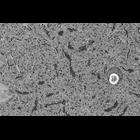
CIL:40261
NCBI Organism Classification
Mus musculus
Biological Process
cerebellum structural organization
Cellular Component
synapse
Single slice of a processed serial block face SEM imaging data set through the molecular layer of the cerebellum of an adult mouse. Data have been converted from 16 bit to 8 bit and low pass filtered...
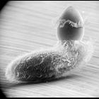
CIL:22782
NCBI Organism Classification
Didinium nasutum
Biological Process
phagocytosis
Cellular Component
oral apparatus
Didinium intiates capture of Paramecium. Believe-it-or-not, this Didinium may accomplish the ingestion of the Paramecium in less than one minute. See other images in the link below. Also showing metac...
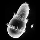
CIL:21992
NCBI Organism Classification
Didinium nasutum
Biological Process
phagocytosis
Cellular Component
oral apparatus
Didinium ingests Paramecium. Note that Paramecium is nearly the waiting food vacuole and the proboscis of Didinium is beginning to reform. This micrograph also shows the metachronous waves of cilia in...
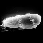
CIL:21995
NCBI Organism Classification
Didinium nasutum
Biological Process
phagocytosis
Cellular Component
oral apparatus
Didinium ingests Paramecium. Note that Paramecium is folded in half as it is compressed and enters the waiting food vacuole. This micrograph also shows a few discharged Paramecium trichocysts as well ...
CIL:39246
NCBI Organism Classification
Didinium nasutum
Biological Process
phagocytosis
Cellular Component
oral apparatus
Didinium ingests Paramecium. At the midpoint of engulfment, some 30 seconds after contact. This micrograph was taken in 1968 by G. Antipa on a Cambridge Mark IIA operating at 20kV. The negative magnif...
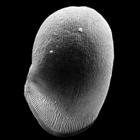
CIL:40906
NCBI Organism Classification
uncultured scuticociliate
Biological Process
cortical cytoskeleton organization
Cellular Component
cilium
Conchophthirus was deciliated by calcium ion shock followed by shearing through a micropipette. This revealed the location of the deciliated basal bodies and the cell's surface architecture. In this i...
CIL:40592
NCBI Organism Classification
Salmonella
Biological Process
induction of bacterial agglutination
Cellular Component
cell surface
This scanning electron micrograph of salmonella bacteria, was made possible by using low kV to visualize the surface detail of the bacteria. The image was taken with the VcD (backscatter detector), u...
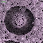
CIL:41473
NCBI Organism Classification
coral
Biological Process
skeleton organization
Cellular Component
calcium skeleton
Scanning electron micrograph of the calcium skeleton of coral polyp. The sample was cleaned with bleach solution, dried and carbon coated prior to imaging. The image was collected using a secondary...
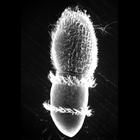
CIL:17892
NCBI Organism Classification
Didinium nasutum
Biological Process
phagocytosis
Cellular Component
oral apparatus
Didinium ingests Paramecium. Also showing a few discharged trichocysts and metachronous waves of cilia in the two characteristic ciliary girdles of Didinium nasutum. This micrograph was taken in 1968 ...
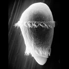
CIL:19537
NCBI Organism Classification
Didinium nasutum
Biological Process
oral apparatus organization
Cellular Component
oral apparatus
The proboscis of Didinium. While not well preserved, this image clearly shows the clockwise surface ridges that are elevated by ribbons of microtubules that are involved in supporting the buccal cavit...
« Previous
1
...
26
27
28
29
30
31
32
33
...
37
Next »
Results per page:
10
20
50
100