Alternate header for print version


Licensing
Public Domain: This image is in the public domain and thus free of any copyright restrictions. However, as is the norm in scientific publishing and as a matter of courtesy, any user should credit the content provider for any public or private use of this image whenever possible. Learn more
tweet  
*CIL – Cell Image Library accession number. Please use this to reference an image.

CIL:19537*  Cite 
Description

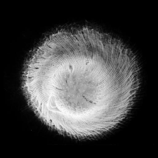
The proboscis of Didinium. While not well preserved, this image clearly shows the clockwise surface ridges that are elevated by ribbons of microtubules that are involved in supporting the buccal cavity during the process of ingesting prey. See the ribbons of microtubules in CIL:17451, 17452, & 15400. The extrusive organelles toxicysts and pexicysts reside just below the membrane of the proboscis plateau. This micrograph was taken in 1968 by G. Antipa on a Cambridge Mark IIA operating at 20kV. The negative magnification is 4760X. The raw film was scanned with an Epson Perfection V750 Pro. This image is available for qualitative analysis. Further details are available at Wessenberg, H. and Antipa, G. 1968. Studies on Didinium nasutum. I. Structure and ultrastructure. Protistologica 4:427-447 and 1970. Capture and ingestion of Paramecium by Didinium nasutum. J. Protozool. 17:250-270.

Biological Sources
NCBI Organism Classification
Didinium nasutum
Cell Type
cell by organism
eukaryotic cell
Eukaryotic Protist
Ciliated Protist
Cellular Component
oral apparatus
microtubule cytoskeleton
cell cortex
cell surface
Biological Context
Biological Process
oral apparatus organization
phagocytosis, engulfment
Attribution
Names
Gregory Antipa (San Francisco State University)
Citation
Digital Object Identifier (DOI)
doi:10.7295/W9CIL19537
Archival Resource Key (ARK)
ark:/b7295/w9cil19537
Grouping This image is part of a group.
Imaging
Image Type
recorded image
Image Mode
illumination by electrons
secondary_electron imaging
Parameters Imaged
illumination by electrons
secondary electron generation
secondary_electron imaging
Source of Contrast
detection of electrons
Visualization Methods
shadowing and plating
Processing History
recorded image
film
Data Qualifiers
raw, unprocessed data
Sample Preparation
Methods
chemically fixed tissue
Parducz fixative
lyophilized specimen
Au/Pd metal sputter-coat
Relation To Intact Cell
whole mounted tissue
Dimensions
Spatial Axis Image Size Pixel Size
X 5835px 3.6nm
Y 4888px 3.6nm