Alternate header for print version
Advanced search
Contributors
Help
Submit
Search
menu
Cell Process
Cell Component
Cell Type
Organism
Microbial
Alzheimer's
Data Sets
University of California, San Diego
9500 Gilman Drive
La Jolla, CA 92093-0608, USA
Voice
: (858) 534-0276
Fax
: (858) 534-7497
Email
: dorloff@ncmir.ucsd.edu
Search Results for
plasma membrane
(608 results)
(Not the results you were expecting?)
(Comments)
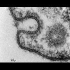
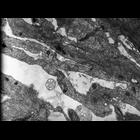
CIL:37137
NCBI Organism Classification
Rattus
Biological Process
regulation of vascular permeability
Cellular Component
clathrin-coated vesicle
Transmission electron micrograph of a clathrin coated pt caveola and transgential cut of a coated vesicle in an endothelial cell. Specimen comes from a capillary in a rat tongue. Image made availabl...
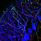
CIL:41920
NCBI Organism Classification
Mus musculus
Biological Process
synaptic connexion organization
Cellular Component
synaptic terminal
Confocal image of synaptic connexions in the mouse olivo-cerebellar system. Purkinje cells (immunofluorescence, blue), olivary axons (anterograde tracing, yellow) and synaptic terminals (immunofluore...
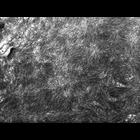
CIL:10051
NCBI Organism Classification
Rana catesbeiana
Biological Process
intermediate filament cytoskeleton organization
Cellular Component
intermediate filament cytoskeleton
This group of micrographs illustrate the ultrastructural characteristics of the middle layer of the three meningeal layers that cover the central nervous system The arachnoid cells contain abundant in...
CIL:10433
NCBI Organism Classification
Rana catesbeiana
Biological Process
intermediate filament cytoskeleton organization
Cellular Component
intermediate filament cytoskeleton
This group of micrographs illustrate the ultrastructural characteristics of the middle layer of the three meningeal layers that cover the central nervous system The arachnoid cells contain abundant in...
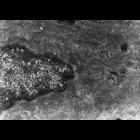
CIL:10439
NCBI Organism Classification
Rana catesbeiana
Biological Process
intermediate filament cytoskeleton organization
Cellular Component
intermediate filament cytoskeleton
This group of micrographs illustrate the ultrastructural characteristics of the middle layer of the three meningeal layers that cover the central nervous system The arachnoid cells contain abundant in...
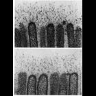
CIL:10931
NCBI Organism Classification
Myotis
Biological Process
extracellular structure organization
Cellular Component
plasma membrane
Filaments extend from microvilli of the epithelial cells in the intestine of the bat, Moytis lucifugus in these two high magnification electron micrographs. These filaments, which are 2.5-5nm thick, ...
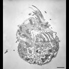
CIL:12320
NCBI Organism Classification
Halteria grandinella
Biological Process
oral apparatus organization
Cellular Component
oral apparatus
This group represents a serial series of sections. At the level of this section, a little deeper in the cell the spongiome of the CV is more pronounced. Oral membranelles in the buccal cavity are indi...
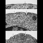
CIL:13003
NCBI Organism Classification
Cavia porcellus
Biological Process
pinocytosis
Cellular Component
coated vesicle
Stages of coated pit vesicle invagination during micropinocytosis. Each frame is a portion of the surface of polychromatophilic erythroblasts from guinea pig bone marrow and various stages of vesicle...
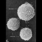
CIL:13091
NCBI Organism Classification
Mus musculus
Biological Process
immune system process
Cellular Component
microvillus
This image shows two different types of white blood cells from a mouse which are essential for mammalian immune response to protect from infection. The larger cell is a macrophage and the smaller two...
CIL:13698
NCBI Organism Classification
Chlorocebus aethiops
Biological Process
G-protein coupled receptor internalization
Cellular Component
plasma membrane
This is one of a group of four images in Figure S5 in Apaja et al., JCB 2010, that support the conclusion that the WT vasopressin type2 receptor (V2R) and the V2R W164S mutant (that causes nephrogenic...
« Previous
1
...
18
19
20
21
22
23
24
25
...
61
Next »
Results per page:
10
20
50
100