Alternate header for print version
Advanced search
Contributors
Help
Submit
Search
menu
Cell Process
Cell Component
Cell Type
Organism
Microbial
Alzheimer's
Data Sets
University of California, San Diego
9500 Gilman Drive
La Jolla, CA 92093-0608, USA
Voice
: (858) 534-0276
Fax
: (858) 534-7497
Email
: dorloff@ncmir.ucsd.edu
Search Results for
plasma membrane
(608 results)
(Not the results you were expecting?)
(Comments)
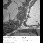
CIL:13132
NCBI Organism Classification
Paramecium multimicronucleatum
Biological Process
cortical cytoskeleton organization
Cellular Component
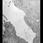
cytoproct
In defecation, when a spent vacuole has fused with the single membrane along the midline of the ridge the lips of the cytoproct split apart and form a wide gap. The membrane of the spent vacuole is no...
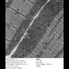
CIL:9813
NCBI Organism Classification
Opercularia [NCBITaxon:168247]
Biological Process
plasma membrane organization
Cellular Component
plasma membrane organization
Image of the pellicle of Opercularia coarctata. Indentations of the plasma membrane penetrate the alveolar membranes end as coated pits. These pits are also known as pellicular pores are homologous to...
CIL:9851
NCBI Organism Classification
Paramecium tetraurelia
Biological Process
microtubule cytoskeleton organization
Cellular Component
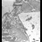
cell cortex
b-tubulin immunogold labeled microtubules arise from the apex of the cytoproct ridge and pass to the spent DV membrane. Trichocyst and trichocyst tip included in this image. TEM taken on 7/5/96 by R. ...
CIL:12312
NCBI Organism Classification
Nassula
Biological Process
contractile vacuole organization
Cellular Component
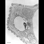
contractile vacuole
Contractile vacuole of Nassula sp. Parts of the single CV appear on two sides of the CV pore. Fields of spongiome abut the filled portions of the CV. TEM taken on 3/27/69 by R. Allen with Philips 300 ...
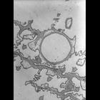
CIL:38894
NCBI Organism Classification
Paramecium caudatum
Biological Process
contractile vacuole organization
Cellular Component
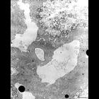
contractile vacuole pore
A circular depression on the dorsal surface of the cell marks the expulsion site of a contractile vacuole. This is referred to as the contractile vacuole “pore.". Microtubules encircle the pore. Tr...
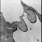
CIL:9807
NCBI Organism Classification
Euplotes sp.
Biological Process
water homeostasis
Cellular Component
contractile vacuole pore
A high resolution image and rare micrograph of the contractile vacuole and intact contractile vacuole membrane of Euplotes. Close examination at high magnification reveals the organization of microtub...
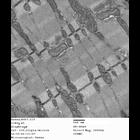
CIL:258
NCBI Organism Classification
Mus musculus
Biological Process
plasma membrane organization
Cellular Component
dystroglycan complex
Thin section electron microscopy of diaphragm (skeletal) muscle from a wild type mouse. Sample was viewed with a Hitachi 7600 electron microscope (accelerating voltage 80 KV) and imaged with an AMT d...
CIL:266
NCBI Organism Classification
Mus musculus
Biological Process
plasma membrane organization
Cellular Component
dystroglycan complex
Thin section electron microscopy of diaphragm (skeletal) muscle from a wild type mouse. Sample was viewed with a Hitachi 7600 electron microscope (accelerating voltage 80 KV) and imaged with an AMT d...
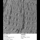
CIL:339
NCBI Organism Classification
Mus musculus
Biological Process
plasma membrane organization
Cellular Component
dystroglycan complex
Thin section electron microscopy of gastrocnemius (skeletal) muscle from a wild type mouse. Sample was viewed with a Hitachi 7600 electron microscope (accelerating voltage 80 KV) and imaged with an A...
CIL:345
NCBI Organism Classification
Mus musculus
Biological Process
plasma membrane organization
Cellular Component
dystroglycan complex
Thin section electron microscopy of gastrocnemius (skeletal) muscle from a wild type mouse. Sample was viewed with a Hitachi 7600 electron microscope (accelerating voltage 80 KV) and imaged with an A...
« Previous
1
...
43
44
45
46
47
48
49
50
...
61
Next »
Results per page:
10
20
50
100