Licensing

Pd
Public Domain: This image is in the public domain and thus free of any copyright restrictions. However, as is the norm in scientific publishing and as a matter of courtesy, any user should credit the content provider for any public or private use of this image whenever possible. Learn more
Comments: Powered by Pnyxe

Description

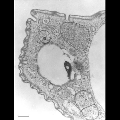
Image of the pellicle of Opercularia coarctata. Indentations of the plasma membrane penetrate the alveolar membranes end as coated pits. These pits are also known as pellicular pores are homologous to parasomal sacs of other ciliates. The surface ridges contain an electron opaque rod in the cytosol of unknown composition, seen here in cross section. TEM taken on 6/11/69 by R. Allen with Philips 300 operating at 60kV. Neg. 30,000X. Bar = 0.25µm. A print of the negative was scanned and processed in Photoshop. This image is best used for qualitative analysis. A high resolution image (CIL:7336) is available for quantitative analysis. Standard glutaraldehyde fixation followed by osmium tetroxide, dehydrated in alcohol and embedded in an epoxy resin. Microtome sections prepared at approximately 75nm thickness. Additional information available at (http://www5.pbrc.hawaii.edu/allen/).

Biological Sources

NCBI Organism Classification
Opercularia [NCBITaxon:168247]
Opercularia coarctata
Cell Type
cell by organism
eukaryotic cell
Eukaryotic Protist
Ciliated Protist
Cell Line
Cambridge Culture Collection, Cambridge, England, # LB1657/1
Cellular Component
GO:0007009
clathrin coat of coated pit
alveolar membranes
rods of unknown material

Biological Context

Biological Process
plasma membrane organization
clathrin coat assembly
cytoskeletal anchoring at plasma membrane
cytoskeletal organization
Molecular Function
clathrin binding

Attribution

Name
Richard Allen (University of Hawaii)
Link
http://www5.pbrc.hawaii.edu/allen/

Grouping

This image is part of a group.

Imaging

Image Type
transmission electron microscopy (TEM)
illumination by electrons
Imaging Mode
detection of electrons
film
Parameters Imaged
electron density
Source of Contrast
stain with broad specificity
Visualization Methods
stain with broad specificity
osmium tetroxide
uranyl salt
lead salt
Processing History
recorded image
film
Print from negative scanned to Photoshop.
Data Qualifiers
processed data
suitable for spatial measurements

Dimensions

Spatial Axis Image Size Pixel Size
X 2753px ——
Y 3624px ——
*CIL – Cell Image Library accession number. Please use this to reference an image.