Alternate header for print version
Advanced search
Contributors
Help
Submit
Search
menu
Cell Process
Cell Component
Cell Type
Organism
Microbial
Alzheimer's
Data Sets
University of California, San Diego
9500 Gilman Drive
La Jolla, CA 92093-0608, USA
Voice
: (858) 534-0276
Fax
: (858) 534-7497
Email
: dorloff@ncmir.ucsd.edu
Search Results for
neuron projection
(712 results)
(Not the results you were expecting?)
(Comments)
CIL:2943
NCBI Organism Classification
Rattus
Biological Process
dendrite development
Cellular Component
microtubule cytoskeleton
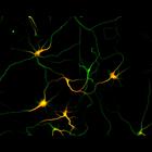
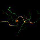
Development of the axon and dendritic arbors in cultured hippocampal neurons after 5 days in vitro. MAP2 staining (red) highlights the dendrites, while microtubule staining (green) reveals both the ax...
CIL:2951
NCBI Organism Classification
Rattus
Biological Process
dendrite development
Cellular Component
microtubule cytoskeleton
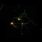
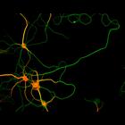
Development of the axon and dendritic arbors in cultured hippocampal neurons after 3 days in vitro. MAP2 staining (red) highlights the dendrites, while microtubule staining (green) reveals both the ax...
CIL:3010
NCBI Organism Classification
Rattus
Biological Process
dendrite development
Cellular Component
microtubule cytoskeleton
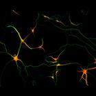
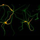
Development of the axon and dendritic arbors in cultured hippocampal neurons after 5 days in vitro. MAP2 staining (red) highlights the dendrites, while microtubule staining (green) reveals both the ax...
CIL:3046
NCBI Organism Classification
Rattus
Biological Process
dendrite development
Cellular Component
microtubule cytoskeleton
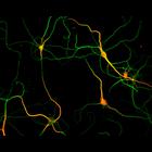
Development of the axon and dendritic arbors in cultured hippocampal neurons after 7 days in vitro. MAP2 staining (red) highlights the dendrites, while microtubule staining (green) reveals both the ax...
CIL:3050
NCBI Organism Classification
Rattus
Biological Process
dendrite development
Cellular Component
microtubule cytoskeleton
Development of the axon and dendritic arbors in cultured hippocampal neurons after 7 days in vitro. MAP2 staining (red) highlights the dendrites, while microtubule staining (green) reveals both the ax...
CIL:3062
NCBI Organism Classification
Rattus
Biological Process
dendrite development
Cellular Component
microtubule cytoskeleton
Development of the axon and dendritic arbors in cultured hippocampal neurons after 7 days in vitro. MAP2 staining (red) highlights the dendrites, while microtubule staining (green) reveals both the ax...
CIL:3070
NCBI Organism Classification
Rattus
Biological Process
dendrite development
Cellular Component
microtubule cytoskeleton
Development of the axon and dendritic arbors in cultured hippocampal neurons after 7 days in vitro. MAP2 staining (red) highlights the dendrites, while microtubule staining (green) reveals both the ax...
CIL:40255
NCBI Organism Classification
Rattus norvegicus
Biological Process
none specified
Cellular Component
mitochondrion
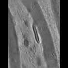
Single computed slice through a tomographic volume of mitochondria at the Node of Ranvier in rat peripheral nerve root. This image has been downsampled from the raw data image which can be accessed us...
CIL:40164
NCBI Organism Classification
Mus musculus
Biological Process
regulation of action potential in neuron
Cellular Component
node of Ranvier
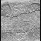
Single slice through a single tilt tomogram of the Node of Ranvier from mouse sciatic nerve. This image has been downsampled from the raw data image which can be accessed using the link provided to th...
CIL:40165
NCBI Organism Classification
Mus musculus
Biological Process
regulation of action potential in neuron
Cellular Component
node of Ranvier
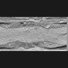
Single slice through a single tilt tomogram of the Node of Ranvier from mouse sciatic nerve prepared by high pressure freezing and freeze substitution of aldehyde fixed material. This image has been ...
« Previous
1
...
18
19
20
21
22
23
24
25
...
72
Next »
Results per page:
10
20
50
100